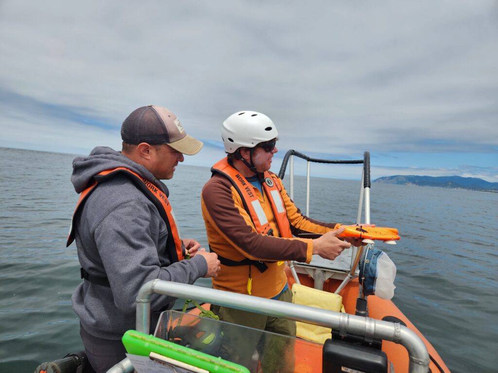
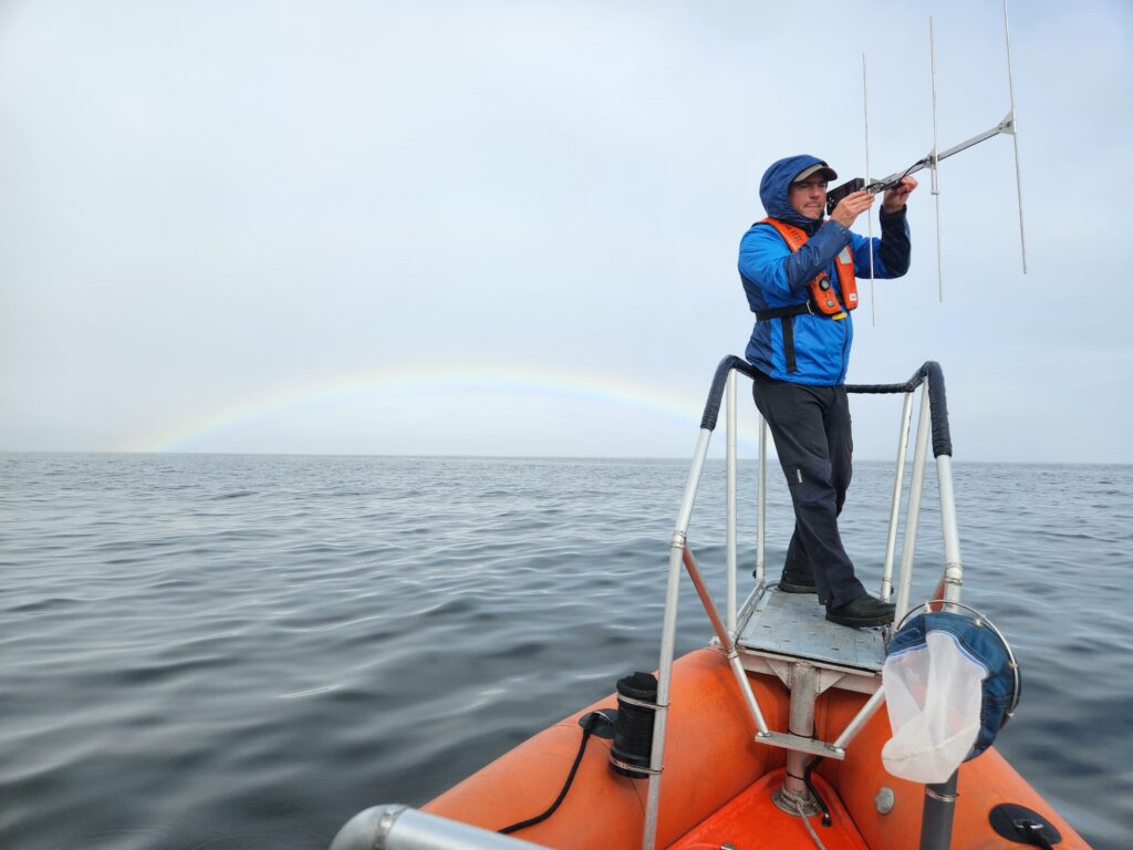
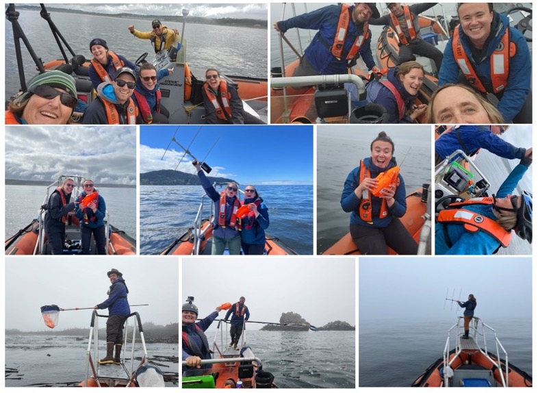
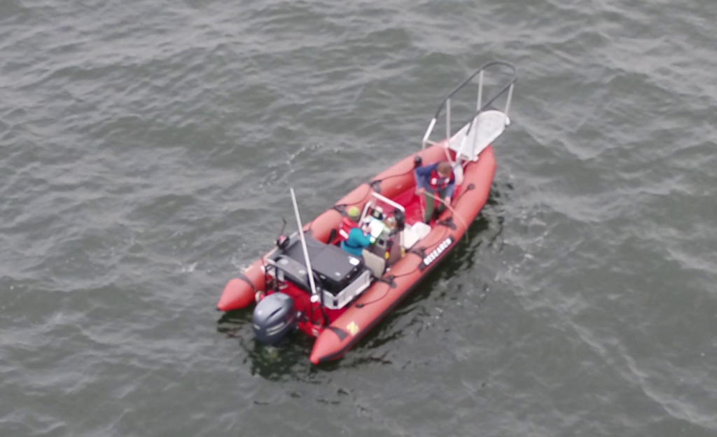
The whales keep coming and we keep learning: a wrap up of the eighth GRANITE field season.

Clara Bird, PhD Candidate, OSU Department of Fisheries, Wildlife, and Conservation Sciences, Geospatial Ecology of Marine Megafauna Lab

As you may remember, last year’s field season was a remarkable summer for our team. We were pleasantly surprised to find an increased number of whales in our study area compared to previous years and were even more excited that many of them were old friends. As we started this field season, we were all curious to know if this year would be a repeat. And it’s my pleasure to report that this season was even better!
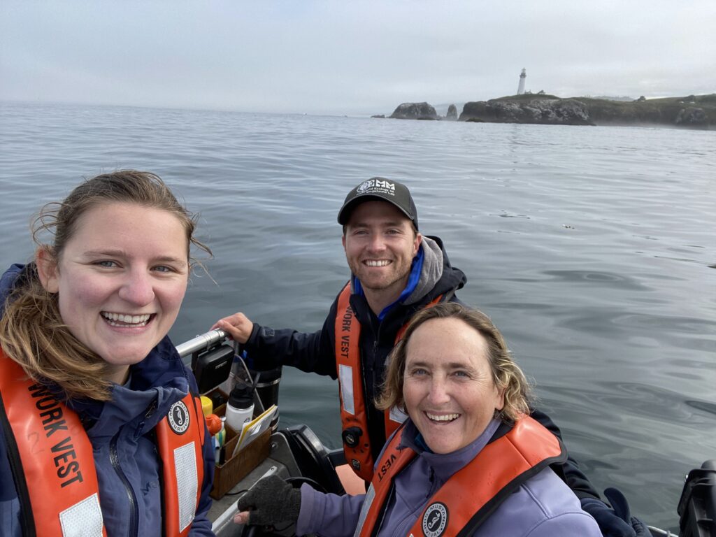
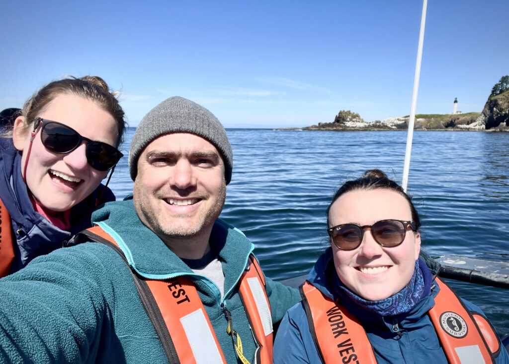
We started the season with an exciting day (6 known whales! see Lisa’s blog) and the excitement (and whales) just kept coming. This season we saw 71 individual whales across 215 sightings! Of those 71, 44 were whales we saw last year, and 10 were new to our catalog, meaning that we saw 17 whales this season that we had not seen in at least two years! There is something extra special about seeing a whale we have not seen in a while because it means that they are still alive, and the sighting gives us valuable data to continue studying health and survival. Another cool note is that 7 of our 12 new whales from last year came back this year, indicating recruitment to our study region.

Included in that group of 7 whales are the two calves from last year! Again, indicating good recruitment of new whales to our study area. We saw both Lunita and Manta (previously nick-named ‘Roly-poly’) throughout this season and we were always happy to see them back in our area and feeding on their own.

Drone image of Lunita from 2023
Drone image of Manta from 2023

We had an especially remarkable encounter with Lunita at the end of this season when we found this whale surface feeding on porcelain crab larvae (video 1)! This is a behavior that we rarely observe, and we’ve never seen a juvenile whale use this behavior before, inspiring questions around how Lunita knew how to perform this behavior.

Not only did we resight our one-year-old friends, but we found two new calves born to well-known mature females (Clouds and Spotlight). We had previously documented Clouds with a calf (Cheetah) in 2016 so it was exciting to see her with a new calf and to meet Cheetah’s sibling! Cheetah has become one of our regulars so we’re curious to see if this new calf joins the regular crew as well. We’re also hoping that Spotlight’s calf will stick around; and we’re optimistic since we observed it feeding alone later in the season.
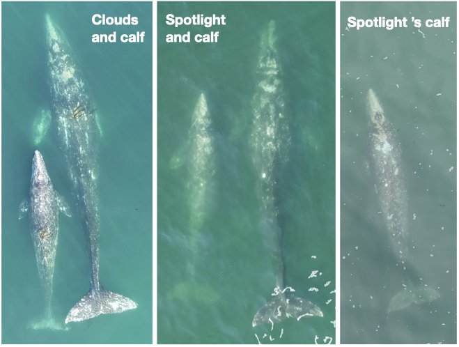
Collage of new calves from 2023! Left: Clouds and her calf, Center: Spotlight and her calf, Right: Spotlight’s calf independently foraging

Of course, 71 whales means heaps of data! We spent 226 hours on the water, conducted 132 drone flights (a record!), and collected 61 fecal samples! Those 132 flights were over 64 individual whales, with Casper and Pacman tying for “best whale to fly over” with 10 flights each. We collected 61 fecal samples from 26 individual whales with a three-way tie for “best pooper” between Hummingbird, Scarlett, and Zorro with 6 fecal samples each. And we continued to collect valuable prey and habitat data through 80 GoPro drops and 79 zooplankton net tows.
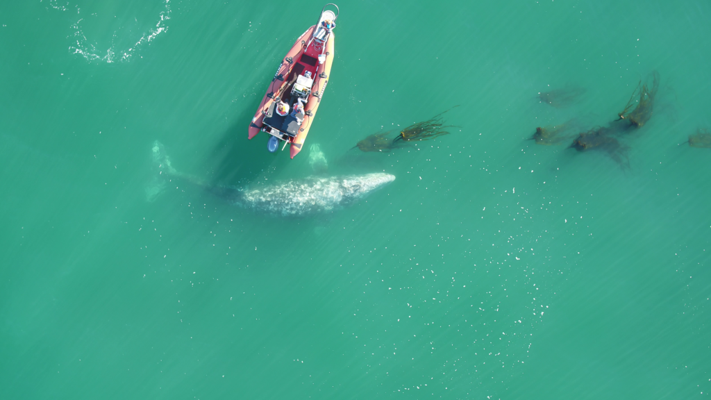
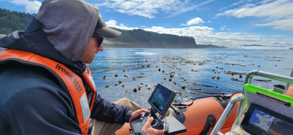
And if you were about to ask, “but what about tagging?!”, fear not! We continued our suction cup tagging effort with a successful window in July where we were joined by collaborators John Calambokidis from Cascadia Research Collective and Dave Cade from Hopkins Marine Station and deployed four suction-cup tags.
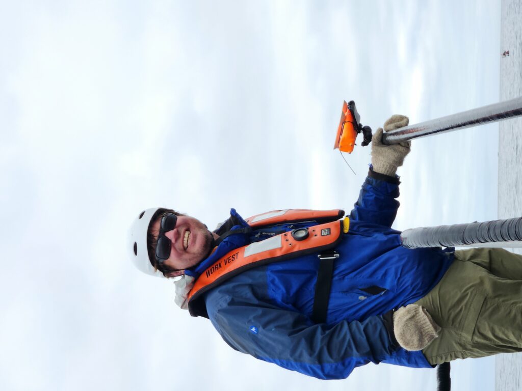
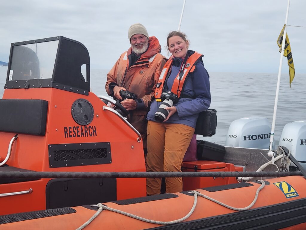
It’s hard to believe all the work we’ve accomplished in the past five months, and I continue to be honored and proud to be on this incredible team. But as this season has come to a close, I have found myself reflecting on something else. Learning. Over the past several years we have learned so much about not only these whales in our study system but about how to conduct field work. And while learning is continuous, this season in particular has felt like an exciting time for both. In the past year our group has published work showing that we can detect pregnancy in gray whales using fecal samples and drone imagery (Fernandez Ajó et al., 2023), that PCFG gray whales are shorter and smaller than ENP whales (Bierlich et al., 2023), and that gray whales are consuming high levels of microplastics (Torres et al., 2023). We also have several manuscripts in review focused on our behavior work from drones and tags. While this information does not directly affect our field work, it does mean that while we’re observing these whales live, we better understand what we’re observing and we can come up with more specific, in-depth questions based on this foundation of knowledge that we’re building. I have enjoyed seeing our questions evolve each year based on our increasing knowledge and I know that our collaborative, inquisitive chats on the boat will only continue inspiring more exciting research.

On top of our gray whale knowledge, we have also learned so much about field work. When I think back to the early days compared to now, there is a stark difference in our knowledge and our confidence. We do a lot on our little boat! And so many steps that we once relied on written lists to remember to do are now just engrained in our minds and bodies. From loading the boat, to setting up at the dock, to the go pro drops, fecal collections, drone operations, photo taking, and photo ID, our team has become quite the well-oiled machine. We were also given the opportunity to reflect on everything we’ve learned over the past years when it was our turn to train our new team member, Nat! Nat is a new PhD student in the GEMM lab who is joining team GRANITE. Teaching her all the ins and outs of our fieldwork really emphasized how much we ourselves have learned.
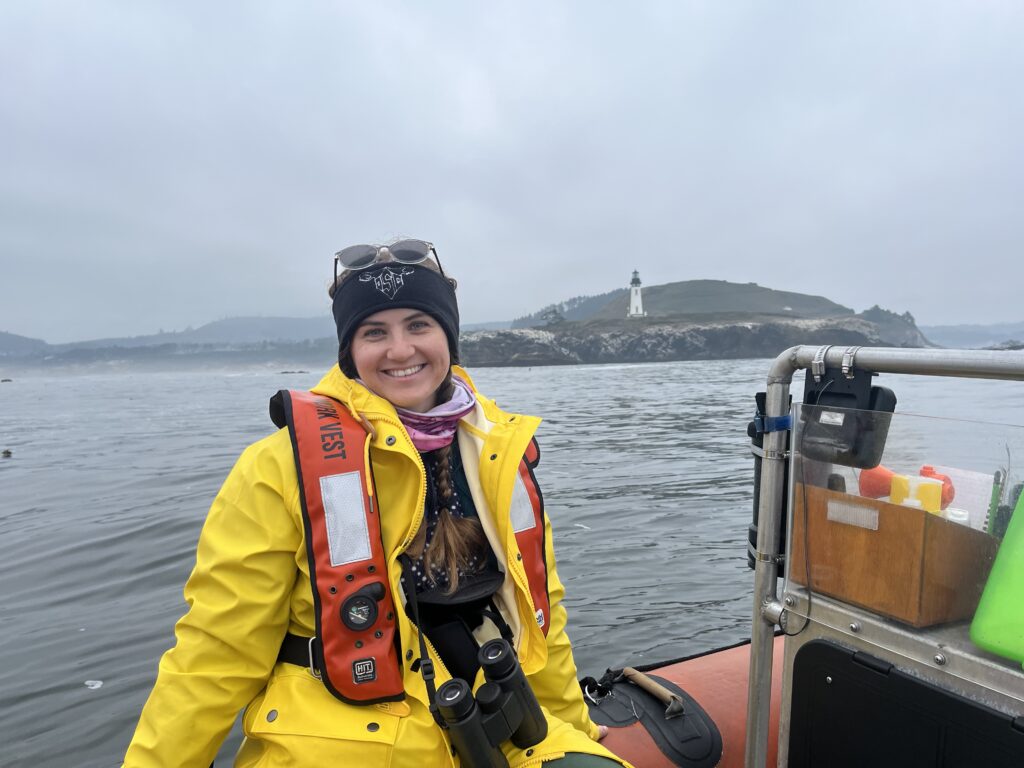
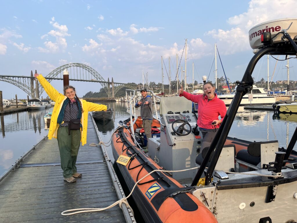
On a personal note, this was my third season as a drone pilot, and honestly, I was pleasantly surprised by my experience this season. Since I started piloting, I have experienced pretty intense nerves every time I’ve flown the drone. From stress dreams, to mild nausea, and an elevated heart rate, flying the drone was something that I didn’t necessarily look forward to. Don’t get me wrong – it’s incredibly valuable data and a privilege to watch the whales from a bird’s eye view in real time. But the responsibility of collecting good data, while keeping the drone and my team members safe was something that I felt viscerally. And while I gained confidence with every flight, the nerves were still as present as ever and I was starting to accept that I would never be totally comfortable as a pilot. Until this season, when the nerves finally cleared, and piloting became as innate as all the other field work components. While there are still some stressful moments, the nerves don’t come roaring back. I have finally gone through enough stressful situations to not be fazed by new ones. And while I am fully aware that this is just how learning works, I write this reflection as a reminder to myself and anyone going through the process of learning any new skill to push through that fear. Remember there can be a disconnect between the time when you know how to do something well, or well-enough, and the time when you feel comfortable doing it. I am just as proud of myself for persevering as I am of the team for collecting so much incredible data. And as I look ahead to my next scary challenge (finishing my PhD!), this is a feeling that I am trying to hold on to. 

Stay tuned for updates from team GRANITE!

Did you enjoy this blog? Want to learn more about marine life, research, and conservation? Subscribe to our blog and get a weekly alert when we make a new post! Just add your name into the subscribe box below!

Loading

References

Bierlich, K. C., Kane, A., Hildebrand, L., Bird, C. N., Fernandez Ajo, A., Stewart, J. D., Hewitt, J., Hildebrand, I., Sumich, J., & Torres, L. G. (2023). Downsized: Gray whales using an alternative foraging ground have smaller morphology. Biology Letters19(8), 20230043. https://doi.org/10.1098/rsbl.2023.0043

Fernandez Ajó, A., Pirotta, E., Bierlich, K. C., Hildebrand, L., Bird, C. N., Hunt, K. E., Buck, C. L., New, L., Dillon, D., & Torres, L. G. (2023). Assessment of a non-invasive approach to pregnancy diagnosis in gray whales through drone-based photogrammetry and faecal hormone analysis. Royal Society Open Science10(7), 230452. https://doi.org/10.1098/rsos.230452

Torres, L. G., Brander, S. M., Parker, J. I., Bloom, E. M., Norman, R., Van Brocklin, J. E., Lasdin, K. S., & Hildebrand, L. (2023). Zoop to poop: Assessment of microparticle loads in gray whale zooplankton prey and fecal matter reveal high daily consumption rates. Frontiers in Marine Science10. https://www.frontiersin.org/articles/10.3389/fmars.2023.1201078

A Gut Feeling: DNA Metabarcoding Gray Whale Diets

By Charles Nye, graduate student, OSU Department of Fisheries, Wildlife, & Conservation Sciences, Cetacean Conservation and Genomics Laboratory

Figure 1: An illustration (by me) of a feeding gray whale whose caudal end transitions into a DNA double helix.

Let’s consider how much stuff organisms shed daily. If you walk down a hallway, you’ll leave a microscopic trail of skin cells, evaporated sweat, and even more material if you so happen to sneeze or cough (as we’ve all learned). The residency of these bits and pieces in a given environment is on the order of days, give or take (Collins et al. 2018). These days, we can extract, amplify, and sequence DNA from leftover organismal material in environments (environmental DNA; eDNA), stomach contents (dietary DNA, dDNA), and other sources (Sousa et al. 2019; Chavez et al. 2021).

You might be familiar with genetic barcoding, where scientists are able to use documented and annotated pieces of a genome to identify a piece of DNA down to a species. Think of these as genetic fingerprints from a crime scene where all (described) species on Earth are prime suspects. With advancements in computing technology, we can barcode many species at the same time—a process known as metabarcoding. In short, you can now do an ecosystem-wide biodiversity survey without even needing to see your species of interest (Ficetola et al. 2008; Chavez et al. 2021).

(Before you ask: yes, people have tried sampling Loch Ness and came up with not a single strand of plesiosaur DNA (University of Otago, 2019).)

I received my crash course on metabarcoding when I was employed at the Monterey Bay Aquarium Research Institute (MBARI), right before grad school. There, I was employed to help refine eDNA survey field and laboratory methods (in addition to some cool robot stuff). Here at OSU, I use metabarcoding to research whale ecology, detection, and even a little bit of forensics  work. Cetacean species (or evidence thereof) I’ve worked on include North Atlantic right whales (Eubalaena glacialis), killer whales (Orcinus spp.), and gray whales (Eschrichtius robustus).

Long-time readers of the GEMM Lab Blog are probably quite knowledgeable about the summertime grays—the Pacific Coast Feeding Group (PCFG). All of us here at OSU’s Marine Mammal Institute (MMI) are keenly interested in understanding why these whales hang out in the Pacific Northwest during the summer months and what sets them apart from the rest of the Eastern North Pacific gray whale population. What interests me? Well, I want to double-check what they’re eating—genetically.

“What does my study species eat?” is a straightforward but underappreciated question. It’s also deceptively difficult to address. What if your species live somewhere remote or relatively inaccessible? You can imagine this is a common logistical issue for most research in marine sciences. How many observations do you need to make to account for seasonal or annual changes in prey availability? Do all individuals in your study population eat the same thing? I certainly like to mix and match my diet.

Gray whale foraging ecology has been studied comprehensively over the last several decades, including an in-depth stomach content evaluation by Mary Nerini in 1984 and GEMMer Lisa Hildebrand’s MSc research. PCFG whales seem to prefer shrimpy little creatures called mysids, along with Dungeness crab (Cancer magister) larvae, during their stay in the Pacific Northwest (PNW), most notably the mysid Neomysis rayii (Guerrero 1989; Hildebrand et al. 2021). Indeed, the average energetic values of common suspected prey species in PNW waters rival the caloric richness of Arctic amphipods (Hildebrand et al. 2021). However, despite our wealth of visual foraging observations, metabarcoding may add an additional layer of resolution. For example, the ocean sunfish (Mola mola) was believed to exclusively forage on gelatinous zooplankton, but a metabarcoding approach revealed a much higher diversity of prey items, including other bony fishes and arthropods (Sousa et al. 2016).

Given all this exposition, you may be wondering: “Charles—how do you intend on getting dDNA from gray whales? Are you going to cut them open?”

Figure 2: The battle station, a vacuum pump that I use to filter out all of the particulate matter from a gray whale dDNA sample. The filter is made of polycarbonate track etch material, which melts away in the DNA extraction process—quite handy, indeed!

No. I’m going to extract DNA from their poop.

Well, actually, I’ve been doing that for the last two years. My lab (Cetacean Conservation and Genomics Laboratory, CCGL) and GEMM Lab have been collaborating to make lemonade out of, er…whale poop. An archive of gray whale fecal samples (with ongoing collections every field season) originally collected for hormone analyses presented itself with new life—the genomics kind. In addition to community-level data, we are also able to recover informative DNA from the gray whales, including sex ID from “depositing” individuals, though the recovery rate isn’t perfect.

Because the GEMM Lab/MMI can non-invasively collect multiple samples from the same individuals over time, dDNA metabarcoding is a great way to repeatedly evaluate the diets of the PCFG, just shy of being at the right place at the right time with a GoPro or drone to witness a feeding event.  While we can get stomach contents and even usable dDNA from a naturally deceased whale, those data may not be ideal. How representative a stranded whale is of the population is dependent on the cause of death; an emaciated or critically injured individual, for example, is a strong outlier.

Figure 3: Presence/absence of the top 10 most-common taxonomic Families observed in the PCFG gray whale dDNA dataset (n = 20, randomly selected). Filled-in dots indicate at least one genetic read associated with that Family, and empty dots indicate none. Note the prey taxa: mysids (Mysidae), krill (Euphausiidae), and olive snails (Olividae).

Here’s a snapshot of progress to date for this dDNA metabarcoding project. I pulled out twenty random samples from my much larger working dataset (n = 82) for illustrative purposes (and legibility). After some bioinformatic wizardry, we can use a presence/absence approach to get an empirical glimpse at what passes through a PCFG gray whale. While I am able to recover species-level information, using higher-level taxonomic rankings summarizes the dataset in a cleaner fashion (and also, not every identifiable sequence resolves to species).

The title of most commonly observed prey taxa belongs to our friends, the mysids (Mysidae). Surprisingly, crabs and amphipods are not as common in this dataset, instead losing to krill (Euphausiidae) and olive snails (Olividae). The latter has been found in association with gray whale foraging grounds but not documented in a prey study (Jenkinson 2001). We also get an appreciable amount of interference from non-prey taxa, most notably barnacles (Balanidae), with an honorable mention to hydrozoans (Clytiidae, Corynidae). While easy to dismiss as background environmental DNA, as gray whales do forage at the benthos, these taxa were physically present and identifiable in Nerini’s (1984) gray whale stomach content evaluation.

So—can we conclude that barnacles and hydrozoans are an important part of a gray whale’s diet, as much as mysids? From decades of previous observations, we might say…probably not. Gray whales are actively targeting patches of crabby, shrimpy zooplankton things, and even employ novel foraging strategies to do so (Newell & Cowles 2006; Torres et al. 2018). However, the sheer diversity of consumed species does present additional dimensionality to our understanding of gray whale ecology.

The whales are eating these ancillary organisms, whether they intend to or not, and this probably does influence population dynamics, recruitment, and succession in these nearshore benthic habitats. After all, the shallow pits that gray whales leave behind post-feeding provide a commensal trophic link with other predatory taxa, including seabirds and groundfish (Oliver & Slattery 1985). Perhaps the consumption of these collateral species affects gray whale energetics and reflects on their “performance”?

I hope to address all of this and more in some capacity with my published work and graduate chapters. I’m confident to declare that we can document diet composition of PCFG whales using dDNA metabarcoding, but what comes next is where one can get lost in the sea(weeds). How does the diet of individuals compare to one another? What about at differing time points? Age groups? How many calories are in a barnacle? No need to fret—this is where the fun begins!

References

Chavez F, Min M, Pitz K, Truelove N, Baker J, LaScala-Grunewald D, Blum M, Walz K,

Nye C, Djurhuus A, et al. 2021. Observing Life in the Sea Using Environmental

DNA Oceanog. 34(2):102–119. doi:10.5670/oceanog.2021.218.

Collins R, Wangensteen OS, O’Gorman EJ, Mariani S, Sims DW, Genner M. 2018. Persistence

of environmental DNA in marine systems. Comm Biol. 1(185).

https://doi.org/10.1038/s42003-018-0192-6

Ficetola GF, Miaud C, Pompanon F, Taberlet P. 2008. Species detection using

environmental DNA from water samples. Biol Lett. 4(4):423–425.

doi:10.1098/rsbl.2008.0118.

Hildebrand L, Bernard KS, Torres LG. 2021. Do Gray Whales Count Calories?

Comparing Energetic Values of Gray Whale Prey Across Two Different Feeding

Grounds in the Eastern North Pacific. Front Mar Sci. 8:683634.

doi:10.3389/fmars.2021.683634.

Jenkinson R. 2001. Gray whale (Eschrichtius robustus) prey availability and feeding ecology in

northern California, 1999-2000 [thesis]. California State Polytechnic University,

Humboldt. 81 p.

Newell CL, Cowles TJ. 2006. Unusual gray whale Eschrichtius robustus feeding in the summer

of 2005 off the central Oregon Coast. Geophys Res Lett. 33(22):L22S11.

doi:10.1029/2006GL027189.

Oliver JS, Slattery PN. 1985. Destruction and Opportunity on the Sea Floor: Effects of

Gray Whale Feeding. Ecology. 66(6):1965–1975. doi:10.2307/2937392.

Sousa LL, Silva SM, Xavier R. 2019. DNA metabarcoding in diet studies: Unveiling

ecological aspects in aquatic and terrestrial ecosystems. Environmental DNA.

1(3):199–214. doi:10.1002/edn3.27.

Sousa LL, Xavier R, Costa V, Humphries NE, Trueman C, Rosa R, Sims DW, Queiroz N.

2016. DNA barcoding identifies a cosmopolitan diet in the ocean sunfish. Sci

Rep. 6(1):28762. doi:10.1038/srep28762.

Torres LG, Nieukirk SL, Lemos L, Chandler TE. 2018. Drone Up! Quantifying Whale Behavior

From a New Perspective Improves Observational Capacity. Front Mar Sci. 5:319.

doi:10.3389/fmars.2018.00319.

University of Otago. 2019. First eDNA study of Loch Ness points to something fishy.

https://www.otago.ac.nz/news/news/otago717609.html. [accessed 2023 Apr 25]

Return of the whales: The GRANITE 2022 field season comes to a close

Clara Bird, PhD Candidate, OSU Department of Fisheries, Wildlife, and Conservation Sciences, Geospatial Ecology of Marine Megafauna Lab

It’s hard to believe that it’s already been four and half months since we started the field season (check out Lisa’s blog for a recap of where we began), but as of this weekend the GRANITE project’s 8th field season has officially ended! As the gray whales wrap up their foraging season and start heading south for the winter, it’s time for us to put our gear into storage, settle into a new academic year, and start processing the data we spent so much time collecting.

The field season can be quite an intense time (40 days equaling over 255 hours on the water!), so we often don’t take a moment to reflect until the end. But this season has been nothing short of remarkable. As you may remember from past blogs, the past couple years (2020-21) have been a bit concerning, with lower whale numbers than previously observed. Since many of us started working on the project during this time, most of us were expecting another similar season. But we were wrong in the best way. From the very first day, we saw more whales than in previous years and we identified whales from our catalog that we hadn’t seen in several years.

Image 1: Collage of photos from our field season.

We identified friends – old and new!

This season we had 224 sightings of 63 individual whales. Of those 63, 51 were whales from our catalog (meaning we have seen them in a previous season). Of these 51 known whales, we only saw 20 of them last year! This observation brings up interesting questions such as, where did most of these whales forage last year? Why did they return to this area this year? And, the classic end of season question, what’s going to happen next year?

We also identified 12 whales that were not in our catalog, making them new to the GEMM lab. Two of our new whales are extra exciting because they are not just new to us but new to the population; we saw two calves this year! We were fortunate enough to observe two mom-calf pairs in July. One pair was of a “new” mom in our catalog and her calf. We nicknamed this calf “Roly-poly” because when we found this mom-calf pair, we recorded some incredible drone footage of “roly-poly” continuously performing body rolls while their mom was feeding nearby (video 1). 

Video 1: “Roly-poly” body rolling while their mom headstands. NOAA/NMFS permit #21678.

The other pair includes a known GEMM lab whale, Luna, and her calf (currently nicknamed “Lunita”). We recently found “Lunita” feeding on their own in early October (Image 2), meaning that they are now independent from its mom (for more on mom-calf behavior check out Celest’s recent blog). We’ll definitely be on the lookout for Roly-Poly and Lunita next year!

Image 2: (left) drone image of Luna and Lunita together in July and (right) drone image of Lunita on their own in October.  NOAA/NMFS permit #21678.

We flew, we scooped, we collected heaps of data!

From our previous blogs you probably know that in addition to photo-ID images, our other two most important forms of data collection are drone flights (for body condition and behavior data) and fecal samples (for hormone analysis). And this season was a success for both! 

We conducted 124 flights over 49 individual whales. The star of these flights was a local favorite Scarlett who we flew over 18 different times. These repeat samples are crucial data for us because we use them to gain insight into how an individual’s body condition changes throughout the season. We also recorded loads of behavior data, collecting footage of different foraging tactics like headstanding, side-swimming, and surfacing feeding on porcelain crab larvae (video 2)!

Video 2: Two whales surface feeding on porcelain crab larvae. NOAA/NMFS permit #21678.

We also collected 61 fecal samples from 26 individual whales (Image 3). The stars of that dataset were Soléand Peak who tied with 7 samples each. These hard-earned samples provide invaluable insight into the physiology and stress levels of these individuals and are a crucial dataset for the project.

Image 3: Photos of fecal sample collection. Left – a very heavy sample, center: Lisa and Enrico after collecting the first fecal sample of the season, right: Clara and Lisa celebrating a good fecal sample collection.

On top of all that amazing data collection we also collected acoustic data with our hydrophones, prey data from net tows, and biologging data from our tagging efforts. Our hydrophones were in the water all summer recording the sounds that the whales are exposed to, and they were successfully recovered just a few weeks ago (Image 4)! We also conducted 69 net tows to sample the prey near where the whales were feeding and identify which prey the whales might be eating (Image 5). Lastly, we had two very successful tagging weeks during which we deployed (and recovered!) a total of 9 tags, which collected over 30 hours of data (Image 6; check out Kate’s blog for more on that).

Image 4 – Photos from hydrophone recovery.
Image 5: Photos from zooplankton sampling.
Image 6: Collage of photos from our two tagging efforts this season.

Final thoughts

All in all, it’s been an incredible season. We’ve seen the return of old friends, collected lots of awesome data, and had some record-breaking days (28 whales in one day!). As we look toward the analysis phase of the year, we’re excited to dig into our eight-year dataset and work to understand what might explain the increase in whales this year.

To end on a personal note, looking through photos to put in this blog was the loveliest trip down memory lane (even though it only ended a few days ago) – I am so honored and proud to be a part of this team. The work we do is hard; we spend long hours on a small boat together and it can be a bit grueling at times. But, when I think back on this season, my first thoughts are not of the times I felt exhausted or grumpy, but of all the joy we felt together. From the incredible whale encounters to the revitalizing snacks to the off-key sing alongs, there is no other team I would rather do this work with, and I so look forward to seeing what next season brings. Stay tuned for more updates from team GRANITE!

Did you enjoy this blog? Want to learn more about marine life, research, and conservation? Subscribe to our blog and get a weekly alert when we make a new post! Just add your name into the subscribe box below!

Loading

The learning curve never stops as the GRANITE project begins its seventh field season

Clara Bird, PhD Student, OSU Department of Fisheries, Wildlife, and Conservation Sciences, Geospatial Ecology of Marine Megafauna Lab

When I thought about what doing fieldwork would be like, before having done it myself, I imagined that it would be a challenging, but rewarding and fun experience (which it is). However, I underestimated both ends of the spectrum. I simultaneously did not expect just how hard it would be and could not imagine the thrill of working so close to whales in a beautiful place. One part that I really did not consider was the pre-season phase. Before we actually get out on the boats, we spend months preparing for the work. This prep work involves buying gear, revising and developing protocols, hiring new people, equipment maintenance and testing, and training new skills. Regardless of how many successful seasons came before a project, there are always new tasks and challenges in the preparation phase.

For example, as the GEMM Lab GRANITE project team geared up for its seventh field season, we had a few new components to prepare for. Just to remind you, the GRANITE (Gray whale Response to Ambient Noise Informed by Technology and Ecology) project’s field season typically takes place from June to mid-October of each year. Throughout this time period the field team goes out on a small RHIB (rigid hull inflatable boat), whenever the weather is good enough, to collect photo-ID data, fecal samples, and drone imagery of the Pacific Coast Feeding Group (PCFG) gray whales foraging near Newport, OR, USA. We use the data to assess the health, ecology and population dynamics of these whales, with our ultimate goal being to understand the effect of ambient noise on the population. As previous blogs have described, a typical field day involves long hours on the water looking for whales and collecting data. This year, one of our exciting new updates is that we are going out on two boats for the first part of the field season and starting our season 10 days early (our first day was May 20th). These updates are happening because a National Science Foundation funded seismic survey is being conducted within our study area starting in June. The aim of this survey is to assess geophysical structures but provides us with an opportunity to assess the effect of seismic noise on our study group by collecting data before, during, and after the survey. So, we started our season early in order to capture the “before seismic survey” data and we are using a two-boat approach to maximize our data collection ability.

While this is a cool opportunistic project, implementing the two-boat approach came with a new set of challenges. We had to find a second boat to use, buy a new set of gear for the second boat, figure out the best way to set up our gear on a boat we had not used before, and update our data processing protocols to include data collected from two boats on the same day. Using two boats also means that everyone on the core field team works every day. This core team includes Leigh (lab director/fearless leader), Todd (research assistant), Lisa (PhD student), Ale (new post-doc), and me (Clara, PhD student). Leigh and Todd are our experts in boat driving and working with whales, Todd is our experienced drone pilot, I am our newly certified drone pilot, and Lisa, Ale, and myself are boat drivers. Something I am particularly excited about this season is that Lisa, Ale, and I all have at least one field season under our belts, which means that we get to become more involved in the process. We are learning how to trailer and drive the boats, fly the drones, and handling more of the post-field work data processing. We are becoming more involved in every step of a field day from start to finish, and while it means taking on more responsibility, it feels really exciting. Throughout most of graduate school, we grow as researchers as we develop our analytical and writing skills. But it’s just as valuable to build our skillset for field work. The ocean conditions were not ideal on the first day of the field season, so we spent our first day practicing our field skills.

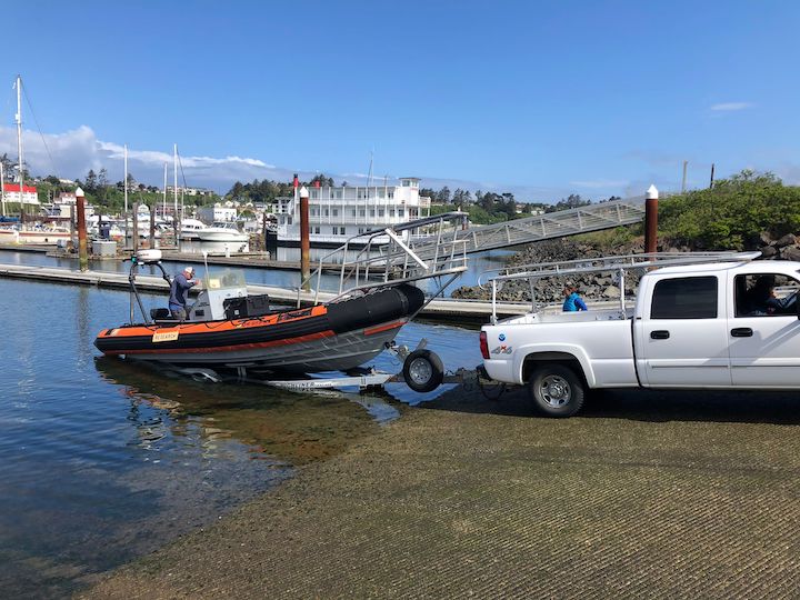
For our “dry run” of a field day, we went through the process of a typical day, which mostly involved a lot of learning from Leigh and Todd. Lisa practiced her trailering and launching of the boat (figure 1), Ale and Lisa practiced driving the boat, and I practiced flying the drone (figure 2). Even though we never left the bay or saw any whales, I thoroughly enjoyed our dry run. It was useful to run through our routine, without rushing, to get all the kinks out, and it also felt wonderful to be learning in a supportive environment. Practicing new skills is stressful to say the least, especially when there is expensive equipment involved, and no one wants to mess up when they’re being watched. But our group was full of support and appreciation for the challenges of learning. We cheered for successful boat launchings and dockings, and drone landings. I left that day feeling good about practicing and improving my drone piloting skills, full of gratitude for our team and excited for the season ahead.

Figure 1. Lisa (driving the truck) launching the boat.
Figure 2. Clara (seated, wearing a black jacket) landing the drone in Ale’s hands.

All the diligent prep work paid off on Saturday with a great first day (figure 3). We conducted five GoPro drops (figure 4), collected seven fecal samples from four different whales (figure 5), and flew four drone flights over three individuals including our star from last season, Sole. Combined, we collected two trifectas (photo-ID images, fecal samples, and drone footage)! Our goal is to get as many trifectas as possible because we use them to study the relationship between the drone data (body condition and behavior) and the fecal sample data (hormones). We were all exhausted after 10 hours on the water, but we were all very excited to kick-start our field season with a great day.

Figure 3. Lisa on the bow pulpit during our first sighting of the day.
Figure 4. Lisa doing a GoPro drop, she’s lowering the GoPro into the water using the line in her hands.
Figure 5. Clara and Ale collecting a fecal sample.

On Sunday, just one boat went out to collect more data from Sole after a rainy morning and I successfully flew over her from launching to landing! We have a long season ahead, but I am excited to learn and see what data we collect. Stay tuned for more updates from team GRANITE as our season progresses!

Are bacteria important? What do we get by analyzing microbiomes?

By Leila Lemos, PhD candidate, Fisheries and Wildlife Department, OSU

As previously mentioned in one of Florence’s blog posts, the GEMM Lab holds monthly lab meetings, where we share updates about our research and discuss articles and advances in our field, among other activities.

In a past lab meeting we were asked to bring an article to discuss that had inspired us in the past to conduct research in the marine field or in our current position. I brought to the meeting a literature review regarding methodologies to overcome the challenges of studying conservation physiology in large whales [1]. This article discusses different non-invasive or minimally invasive matrices (e.g., feces, blow, skin/blubber) that can be gathered from whales, and what types of analyses could be carried out, as well as their pros and cons.

One of the possible analyses that can be performed with fecal samples that was discussed in the article is the gut microflora (i.e., bacterial gut community) via genetic analysis. Since my PhD project analyzes fecal samples to determine/quantify stress responses in gray whales, we have since discussed the possibility of integrating this extra parameter to our analysis.

But… what is the importance of analyzing the gut microflora of a whale? What is the relationship between microflora and stress responses? Should we really use our limited sample size, time and money to work on this extra analysis? In order to be able to answer all of these questions, I began reading some articles of the field to better understand its importance and what kind of research questions this analysis can answer.

The gut of a mammal comprises a natural habitat for a large and dynamic community of bacteria [2] that is first developed in early life. Colonization of facultative bacteria (i.e., aerobic bacteria) begins at birth [3], and later, anaerobic bacteria also colonizes the gut. In humans, at the age of 1 year old, the microbiome should have a stable adult-like signature (Fig. 1).

Figure 01: Development of the microbiome in early life.
Source: [3]
 

The gut bacterial community is important for the physiology and pathology of its host and plays an important role in mammal digestion and health [2], responsible for many metabolic activities, including:

  • fermentation of non-digestible dietary residue and endogenous mucus [2];
  • recovery of energy [2];
  • recovery of absorbable nutrients [2];
  • cellulose digestion [4];
  • vitamin K synthesis [4];
  • important trophic effects on intestinal epithelia (cell proliferation and differentiation) [2];
  • angiogenesis promotion [4];
  • enteric nerve function [4];
  • immune structure [2];
  • immune function [2];
  • protection of the colonized host against invasion by alien microbes (barrier effect) [2];

Despite all the benefits, the bacterial community might also be potentially harmful when changes in the community composition (i.e., dysbiosis) occur due to the use of antibiotics, illness, stress, aging, lifestyle, bad dietary habits [4], and prolonged food and water deprivation [5]. Thus, potential pathological disorders might emerge when the microbiome community changes, such as allergy, obesity, diabetes, autism, multisystem organ failure, gastrointestinal and prostate cancers, inflammatory bowel diseases (IBD), and cardiovascular diseases [2, 4].

Changes in gut bacterial composition may also alter the brain-gut axis and the central nervous system (CNS) signaling [3]. More specifically, the core pathway affected is the hypothalamic-pituitary-adrenal (HPA) axis, which is activated by physical/psychological stressors. According to a previous study [6], the microbial community in the gut is critical for the development of an appropriate stress response. In addition, the microbial colonization in early life should occur within a certain time window, otherwise an abnormal development of the HPA axis might happen.

However, the gut microbiome can not only affect the HPA axis, but the opposite can also occur [3]. Signaling molecules released by the axis can alter the gastrointestinal (GIT) environment (i.e., motility, secretion, and permeability) [7]. Stress responses, as well as diseases, may also alter the gut permeability, causing the bacteria to cross the epithelial barrier (reducing the overall numbers of bacteria in the gut), activating immune responses that also alter the composition of the bacterial community in the gut [8, 9].

Figure 02: Communication between the brain, gut and microbiome in a healthily and in a stressed or diseased (mucosal inflammation) mammal.
Source: [3]
 

Thus, when thinking about whales, monitoring of the gut microflora might allow us to detect changes caused by factors such as aging, illness, prolonged food deprivation, and stressful events [2, 5]. However, since these are two-way factors, it is important to find an association between bacterial composition alterations and stressful events, such as the presence of predators (e.g., killer whales), illness (e.g., bad body condition), prolonged food deprivation (e.g., low prey availability and high competition), noise (e.g., noisy vessel traffic, fisheries opening and seismic surveys), and stressful reproductive status (e.g., pregnancy and lactating period). Examination of possible shifts in the gut microflora may be able to detect and be linked to many of these events, and also forecast possible chronic events within the population. In addition, the bacterial community monitoring study could aid in validating the hormone data (i.e., cortisol) we have been working with.

Therefore, the main research questions that arise in this context that can aid in elucidating the stress physiology in gray whales are:

  1. What is the microflora community content in guts of gray whales along the Oregon coast?
  2. Is it possible to detect shifts in the gut microflora from our gray fecal samples over time?
  3. How do gut microflora and cortisol levels correlate?
  4. Am I able to correlate shifts in gut microflora with any of the stressful events listed above?

We can answer so many other questions by analyzing the microbiome of baleen whales. Microbiomes are mainly correlated with host diet [10], so the composition of a microbiome can be associated with specific diets and functional gut capacity, and consequently, be linked to other animal populations, which helps to decode evolutionary questions. Results of a previous study on baleen whale microbiomes [10] point out that whales harbor unique gut microbiomes that are actually similar to those of terrestrial herbivores. Baleen whales and terrestrial herbivores have a shared physical structure of the GIT tract itself (i.e., multichambered foregut) and a shared hole for fermentative metabolisms. The multichambered foregut of baleen whales fosters the maintenance of the gut microbiome that is capable of extracting relatively unavailable nutrients from zooplankton (i.e., chitin, “sea cellulose”).

Figure 03: The similarities between whale and other terrestrial herbivore gut microbiomes: sea and land ruminants.
Source: [11]
 

Thus, the importance of studying the gut microbiome of a baleen whale is clear. Monitoring of the bacterial community and possible shifts can help us elucidate many questions regarding diet, overall health, stress physiology and evolution. Thinking about my PhD project, it may also help in validating our cortisol level results. I am confident that a microbiome analysis would significantly enhance my studies on the health and ecology of gray whales.


References

  1. Hunt, K.E., et al., Overcoming the challenges of studying conservation physiology in large whales: a review of available methods.Conservation Physiology, 2013. 1: p. 1-24.
  2. Guarner, F. and J.-R. Malagelada, Gut flora in health and disease.The Lancet, 2003. 360: p. 512–519.
  3. Grenham, S., et al., Brain–gut–microbe communication in health and disease.Frontiers in Physiology, 2011. 2: p. 1-15.
  4. Zhang, Y., et al., Impacts of Gut Bacteria on Human Health and Diseases.International Journal of Molecular Sciences, 2015. 16: p. 7493-7519.
  5. Bailey, M.T., et al., Stressor exposure disrupts commensal microbial populations in the intestines and leads to increased colonization by Citrobacter rodentium.Infection and Immunity, 2010. 78: p. 1509–1519.
  6. Sudo, N., et al., Postnatal microbial colonization programs the hypothalamic-pituitary-adrenal system for stress response in mice.The Journal of Physiology, 2004. 558: p. 263–275.
  7. Rhee, S.H., C. Pothoulakis, and E.A. Mayer, Principles and clinical implications of the brain–gut–enteric microbiota axis Nature Reviews Gastroenterology & Hepatology, 2009. 6: p. 306–314.
  8. Kiliaan, A.J., et al., Stress stimulates transepithelial macromolecular uptake in rat jejunum.American Journal of Physiology, 1998. 275: p. G1037–G1044.
  9. Dinan, T.G. and J.F. Cryan, Regulation of the stress response by the gut microbiota: Implications for psychoneuroendocrinology.Psychoneuroendocrinology 2012. 37: p. 1369—1378.
  10. Sanders, J.G., et al., Baleen whales host a unique gut microbiome with similarities to both carnivores and herbivores.Nature Communications, 2015. 6(8285): p. 1-8.
  11. El Gamal, A. Of whales and cows: the baleen whale microbiome revealed. Oceanbites 2016[cited 2018 07/31/2018]; Available from: https://oceanbites.org/of-whales-and-cows-the-baleen-whale-microbiome-revealed/.


Challenges of fecal hormone analyses (Round 2): finally in Seattle!

By Leila Lemos, Ph.D. Student, Department of Fisheries and Wildlife, OSU

In a previous blog of mine, you could read about the challenges I have been facing while I am learning to analyze the hormone content in fecal samples of gray whales (Eschrichtius robustus). New challenges appeared along the way over the last month, while I was doing my training at the Seattle Aquarium (Fig. 1).

Figure 1: View of the Seattle Aquarium.


My training lasted a week and I am truly grateful to the energy and time our collaborators Shawn Larson (research coordinator), Amy Green and Angela Smith (laboratory technicians) contributed. They accompanied me throughout my training to ensure I would be able to conduct hormonal analysis in the future, and to handle possible problems along the way.

The first step was weighing all of the fecal samples (Fig. 2A). Subsequently, the samples were transferred to appropriate glass tubes (Figs. 2B & 2C) for the next laboratorial step.

Figure 2: Analytical processes: (A) Sample weighing; (B) Transference of the sample to a glass tube; (C) Result from the performed steps.


The second conducted step was the hormone extraction. The extraction began with the addition of an organic solvent, called methanol (CH3OH), to the sample tubes (Fig. 3A & 3B). Hormones leach out from the samples and dissolve in the methanol, due to their affinity for this polar solvent.

Tubes were then placed on a plate shaker (Fig. 3C) for 30 minutes, which is used to mix the substances, in order extract the hormones from the fecal samples. The next step was to place the tubes in a centrifuge (Fig. 3D) for 20 minutes. The centrifuge uses the sedimentation principle, causing denser substances or particles to settle to the bottom of the tube, while the less dense substances rise to the top.

Figure 3: Analytical processes: (A) Methanol addition; (B) Sample + methanol; (C) Plate shaker; (D) Centrifuge.


After this process, the two different densities were separated: the high-density particles of the feces were in the bottom of the tube, while the methanol containing the extracted hormones was at the top. The top phase (methanol + hormones) was then pipetted into a different tube (Fig. 4A). The solvent was then evaporated, by using an air dryer apparatus (Fig. 4B), with only the hormones remaining in the tube.

The third performed step was dilution. A specific amount of water, measured in correlation with sample weight and to the amount of the methanol mixed with each sample, was added to each tube (Fig. 4C). Since the hormones were concentrated in the methanol, the readings would exceed the measurement limits of the equipment (plate reader). Thus, in order to prepare the extracts for the immunoassays, different dilutions were made.

Figure 4: Analytical processes: (A) Methanol transference; (B) Methanol drying; (C) Water addition.


The fourth and final step was to finally conduct the assays. Each assay kit is specific to the hormone to be analyzed with specified instructions for each kit. Since we were analyzing four different hormones (cortisol, testosterone, progesterone, and triiodothyronine – T3) we followed four different processes accordingly.

First, a table was filled with the identification numbers of the samples to be analyzed in that specific kit (Fig. 5A). The kit (Fig. 5B) includes the plate reader and several solutions that are used in the process to prepare standard curves, to initiate or stop chemical reactions, among other functions.

A standard curve, also known as calibration curve, is a common procedure in laboratory analysis for determining the concentration of an element in an unknown sample. The concentration of the element is determined by comparison with a set of standard samples of known concentration.

The plate contains several wells (Fig. 5C & 5D), which are filled with the samples and/or these other solutions. When the plate is ready, (Fig.5D) it is carried to the microplate reader that measures the intensity of the color of each of the wells. The intensity of the color is inversely proportional to the concentration of the hormone in both the standards and the samples.

Figure 5: (A) Filling the assay table with the samples to be analyzed; (B) Assay kit to be used; (C) Preparation of the plate; (D) Plate ready to be read.


Since this is the first fecal hormone analysis being performed in gray whales, a validation process of the method is required. Two different tests (parallelism and accuracy) were performed with a pool of three different samples. Parallelism tests that the assay is measuring the antigen (hormone) of interest and also identifies the most appropriate dilution factor to be used for the samples. Accuracy tests that the assay measurement of hormone concentration corresponds to the true concentration of the sample (Brown et al. 2005).

This validation process only needs to be done once. Once good parallelism and accuracy results are obtained, and we have identified the correct dilution factor and approximate concentration of the samples, the samples are ready to be analyzed. Below you can see examples of a good parallelism test (parallel displacement; Fig. 6) and bad parallelism tests (Fig. 7) that indicate no displacement, low concentration or non-parallel displacement; and a good accuracy test (Fig. 8).

Figure 6: Example of a good parallelism test. The dark blue line indicates the standard curve; the pink line indicates a good parallelism test, showing a parallel displacement; and the ratios in black indicate the dilution factors.
Source: Brown et al. (2005)


Figure 7: Examples of bad parallelism tests. The dark blue line indicates the standard curve; the light blue line is an example of no displacement; the pink line is an example of low concentration of the sample; and the green line is an example of non-parallel displacement.
Source: Brown et al. (2005)


Figure 8: Example of a good accuracy test while analyzing hormone levels of pregnanediol glucuronide (Pdg) in elephant urine. The graph shows good linearity (R2 of 0.9986) and would allow for accurate concentration calculations.
Source: Brown et al. (2005)


After the validation tests returned reliable results, the samples were also analyzed. However, many complications were encountered during the assay preparations and important lessons were learned that I know will allow this work to proceed more smoothly and quickly in the future. For instance, I now know to try to buy assay kits of the same brand, and to be extremely careful while reading the manual of the process to be performed with the assay kit. With practice over the coming years, my goal is to master these assay preparations.

Now, the next step will be to analyze all of the results obtained in these analyses and start linking the multiple variables we have from each individual, such as age, sex and body condition. The results of this analysis will lead to a better understanding of how reproductive and stress hormones vary in gray whales, and also link these hormone variations to nutritional status and noise events, one of my PhD research goals.


Cited Literature:

Brown J, Walker S and Steinman K. 2005. Endocrine manual for reproductive assessment of domestic and non-domestic species. Smithsonian’s National Zoological Park, Conservation and Research Center, Virginia 1-69.

Challenges of fecal analyses (Round 1)

By Leila Lemos, Ph.D. Student, Department of Fisheries and Wildlife, OSU

Fieldwork is done for the year and lab analyses just started with some challenges. This is not unexpected since no previous hormonal analysis has been conducted with any gray whale tissue, and whale fecal sample analysis is a relatively new technique. So, I have been thinking, learning, consulting, and creating a methodology as I go along. I am grateful to the expert advice and help from many great collaborators:

  • Kathleen Hunt (Northern Arizona University, AZ, United States)
  • Shawn Larson (Seattle Aquarium, WA, United States)
  • Amy Green (Seattle Aquarium, WA, United States)
  • Rachel Ann Hauser-Davis (Fiocruz, RJ, Brazil)
  • Maziet Cheseby (Oregon State University, OR, United States)
  • Scott Klasek (Oregon State University, OR, United States)

I have learned that an important step before undertaking fecal a hormonal analysis is the desalting process of the samples since salts can interfere in hormonal determinations, leading to false results. In order to remove salt content, each sample was first filtered (Fig. 1A), to remove a majority of the salt water content (Fig. 1B) that is inevitably collected along with the fecal sample. Each sample was then re-suspended in ultra-pure water, to dilute the remaining salt content in a higher water volume (Fig. 1C).

Figure 1: Analytical processes: (A) Filtration of the samples; (B) Result from filtration; (C) Addition of pure water to the samples.

After these steps were completed for each sample, the samples were centrifuged (Fig. 2A) to  precipitate the fecal matter and leave the lighter salt ions in the supernatant (the liquid lying above a solid residue; Fig. 2B). After finishing these two phases, the water was removed with aid of a plastic pippete (Fig. 2C), and I was left with only desalted fecal at the bottom of the tubes (Fig. 2D).

Figure 2: Analytical processes: (A) Samples centrifugation; (B) Result from the centrifugation; (C, D) Results from separating water and sample.

The fecal samples were then frozen at -80°C (Fig. 3A & 3B) and then freeze-dried on a lyophilizer for 2 days to remove all remaining water content (Fig. 3C). Finally, I have what I need: desalted, dry fecal samples, ready for hormone analysis (Fig. 3D).

Figure 3: Analytical processes: (A) Freezing process of the samples; (B) Frozen samples ready to go to the lyophilizer; (C) Samples in the lyophilizer; (D) Final result of the lyophilizing process.

Writing this now, this process seems simple, but it was laborious, and took time to find the equipment needed at the right times. The end product is crucial to get a good final result, so my time investment (and my own increased stress level!) was worth it. This type of analysis is very new for marine mammals and our research lab is still in the learning the best methods. Along the way we were unsure of some decisions, some mistakes were made, and we were afraid of losing precious fecal material. But, this is the fun and challenge of working with a new species and new type of sample and, importantly, we have developed a working protocol that should make the process more efficient and reduce our stress levels next time around.

At the end of this sample preparation process, our 53 samples look great and are ready to be analyzed during my training at the Seattle Aquarium. We are also planning to analyze the water that was removed from the samples (Fig. 2D) to see if any hormone leached out from the poop into the water.

Results from this process will aid in future whale fecal hormone studies. Perhaps only the centrifugation step is needed and we can discard the water without losing hormone content. Or, perhaps we need to analyze both portions of the sample and sum the hormones found in each. We shall know the answer when we get our hormone metabolite results. Just another protocol to be worked out as I move ahead with the hormone analysis of these fecal samples. And through all these challenges I keep the end goal of this work in my mind: to learn about the reproductive and stress hormonal variation in gray whales and to link these variations to nutritional status and noise events. Onward!