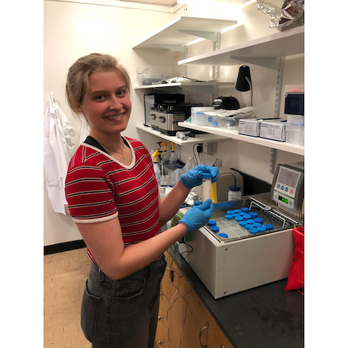
There’s a big difference between human time and Earth–or soil–time. It’s what makes climate impacts so difficult to imagine, and climate solutions so challenging to fully realize. Take it from someone who knows: Adrian Gallo has spent the last decade studying the very idea of “permanence.”
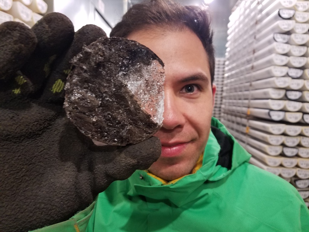
Adrian has dug through a lot of dirt. As a recent PhD graduate in soil science, his research focused on the carbon sequestration potentials of soil. Soil holds about twice as much carbon as our atmosphere. If you factor in permafrost (frozen soils in cold regions that are rapidly thawing) then soil holds nearly three times more carbon than both the atmosphere and all vegetation combined. And that’s a lot.
Let’s back up a second for a quick carbon cycle overview: plants use CO2 to produce sugars through photosynthesis. Microbes eat these sugars, inhaling oxygen and respiring CO2, and when plants and soil decay, they release carbon dioxide back into the atmosphere. There’s a delicate balance between soil being a carbon sink (absorbing more carbon than it releases) or a carbon source (the opposite). More carbon dioxide in the atmosphere = more greenhouse gasses; more climate uncertainty.
Soil’s a tricky thing to study. The age of carbon stored in soil ranges widely. Some plant-derived carbon enters the soil and cycles back into the atmosphere in a number of hours, but other soil carbon can remain underground for thousands of years. And around 12,000 years ago (right around the end of the last ice age) soils used to hold nearly 10% more organic carbon than they do now. Most of that carbon loss came along with the spread of industrial agriculture in the last 200 years. If we could regain some of that carbon storage capacity, we’d have a powerful natural climate solution.
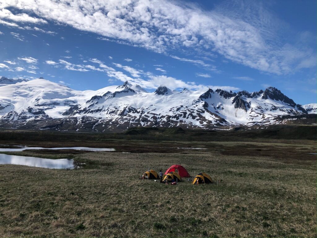
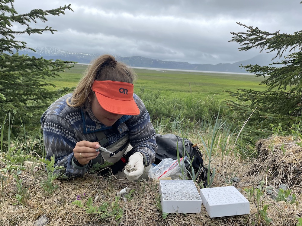
Adrian examined soil cores from nearly 40 representative ecosystems across North America. Adrian’s research was unique in not only its depth (at below 30 cm they tested beyond most existing soil research) but also its length (part of a 30 year project).
The findings? First, soil can indeed be a natural climate solution, but only if farmers can be convinced to alter their land management practices in perpetuity. Many land management practices to prevent carbon escape have been largely the same since the Dust Bowl (minimize tilling, plant natural windbreaks, cover crops, etc) but the expense has not made the switch financially worthwhile. To incentivize farmers, the emerging carbon market allows farm managers to get paid for the carbon they store by selling credits to large companies wanting to offset their emissions. It’s an interesting idea, but also plagued with problems. Big corporations are eager to market themselves as more climate friendly, which often leads to greenwashing. But more importantly, there’s a big question over how long this carbon needs to stay in the soil in order for it to count as a credit. It’s easier to motivate a farmer to alter their land management for 30 years–but that’s thinking in human time, not soil time, and that shortsightedness has some dire consequences, even if moving in the right direction. Now try convincing farmers to use these practices for 100 years–still not on the same scale as soil, but certainly getting closer, and an even tougher sell.
Second, much to Adrian’s and the other researchers’ surprise, there seemed to be a homogenizing effect in endmembers of the soil. No matter what plant types grew aboveground, the distribution of plant-end-members was largely the same, from grasslands to mountain ranges. Adrian coined this term “ecosystem inertia” and it’s still not known why exactly this happens.
This week we have Andrea Domen, a MS student in Food Science and Technology co-advised by Dr. Joy Waite-Cusic and Dr. Jovana Kovacevic, joining us to discuss her research investigating some mischievous pathogenic microbes. Much like an unwelcome dinner guest, food-bourne pathogens can stick around for far longer than you think. Andrea seeks to uncover the mechanisms that allow for Listeria monocytogenes, a ubiquitous pathogen found in dirt that loves cheese (who doesn’t?), to persist in dairy processing facilities.
Listeria hysteria
Way back in the early 2000s, there were two listeriosis outbreaks that were linked to cheese. Because of these two outbreaks, the British Columbia Centre for Disease Control conducted a sampling program over the course of a decade. From this program, 88 isolates of L. monocytogenes from five different facilities were recovered. Within this set of isolates, 63 were from one facility which is now (perhaps unsurprisingly) shut down. Those 63 microbes were essentially clones of each other, which means this one lineage of microbes seemed to carry something that allowed them to survive for multiple years. So how did that lineage of Listeria survive? Turns out, like a 1990’s Reebok, they pump it. Listeria uses a protein in its cell membrane called an efflux pump to remove harmful chemicals like sanitizers, antibiotics, and heavy metals from the cell. Essentially, when the cell absorbs something that is too spicy – it’ll yeet it back out.
Don’t cry over contaminated milk
The idea that food borne pathogens are evolving to withstand processing environments is alarming, but fret not, the results of Andrea’s research are a first step to avoiding the creation of these super microbes in the first place. Instead, it can serve as a warning story for dairy production facilities about what can happen when L. monocytogenes contamination isn’t properly handled. In healthcare, it’s not uncommon to treat a microbial pathogen with multiple medications – as becoming resistant to several treatments is harder for the microbe than becoming resistant to just one. We are also able to apply this treatment method to sanitizing food production facilities by combining different sanitizers – but that is best left up to the chemists to avoid accidentally making an explosion or lethal gas.
To hear more about how Listeria can survive better than Destiny’s Child be sure to listen live on Sunday, May 7th at 7PM on 88.7FM, or download the podcast.
Lots of terrestrial invertebrates have bad reputations. Spiders, bees, flies, wasps, ants. They’re thought of as pests in the garden or they are perceived as threatening, possibly wanting to sting or bite us. I’ll admit it, I’m terrified and grossed out by most invertebrates every time I see one in my house. But this week’s guest may have successfully managed to get me to change my tune…
Scott Mitchell is a 4th year PhD student in the Department of Fisheries, Wildlife, and Conservation Sciences advised by Dr. Sandy DeBano. His overarching research goal is to understand how different land management practices may impact beneficial invertebrate communities in a variety of managed landscapes. Yes, you read that right: beneficial invertebrates. Because while many invertebrates have a bad rep, they’re actually unsung heroes of the world. They pollinate plants, aerate soil, eat actual pest invertebrates and are prey for many other species. In order to tackle his overarching research goal, Scott is conducting two studies in Oregon; one focuses on native bees while the second looks at non-pollinators such as wasps, spiders, and beetles.
(See captions for images at the end of the blog post)
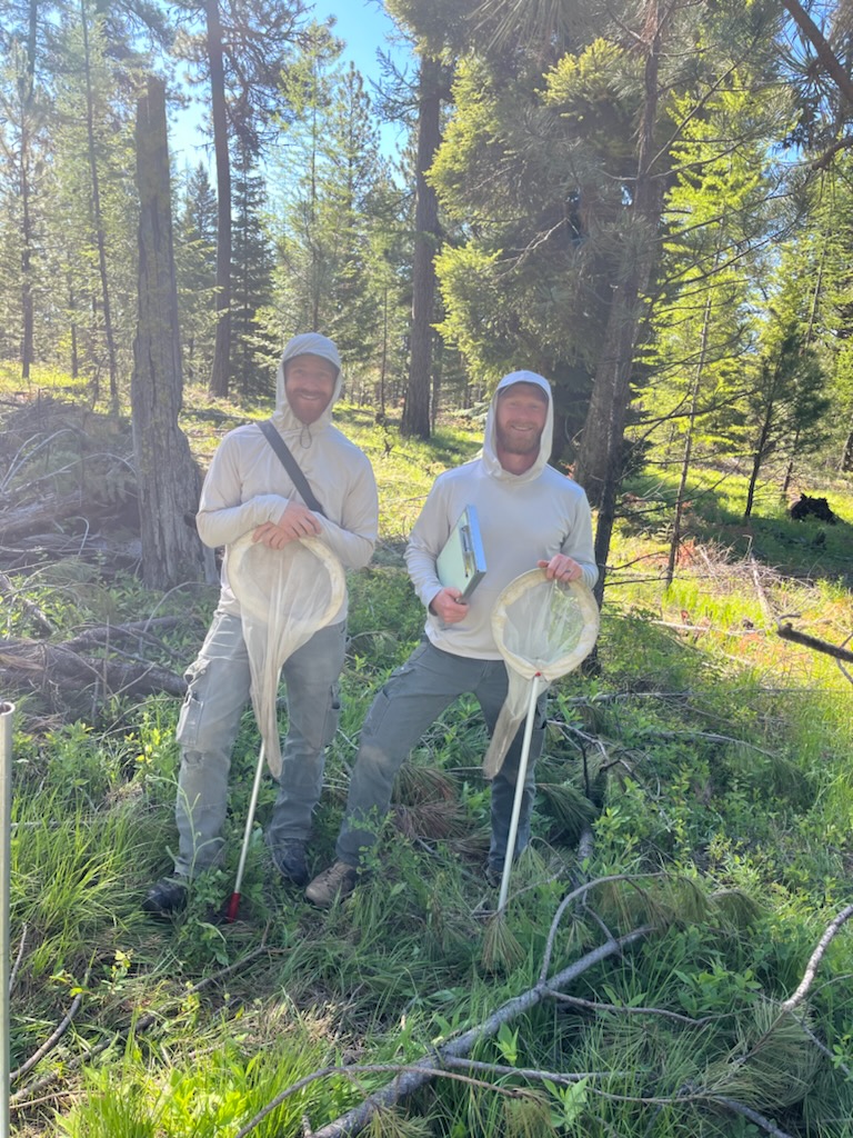
The first study occurs in the Starkey Experimental Forest and Range which is managed by the US Forest Service. The initial research at Starkey in the 1900s was about how cattle grazing impacts on the land. Since then, many more studies have been undertaken and are ongoing, including about forest management, wildlife, plants, and recreation. For Scott’s study, he is collaborating with the Forest Service to look how bee community composition may differ in a number of experimental treatments that are already ongoing at Starkey. The two treatments that Scott is looking into are thinning (thinned vs unthinned forest) and ungulate density (high vs low). The current hypothesis is that in high ungulate densities, flower booms may be reduced due to high grazing and trampling by many ungulate (specifically elk) individuals, thus reducing the number of available blooms to bees. While in the thinning treatments, Scott is expecting to see more flower blooms available to bees in the thinned sites due to increased access to light and resources because of a reduced tree canopy cover. To accomplish this project, Scott collects bee samples in traps and handnets, as well as data on blooming plants.
(See captions for images at the end of the blog post)
Scott’s second study explores non-pollinator community composition in cherry orchards in the Dalles along the Columbia River Gorge. Agricultural landscapes, such as orchards, are heavily managed to produce and maximize a particular agricultural product. However, growers have options about how they choose to manage their land. So, Scott is working closely with a grower to see how different plants planted underneath orchards can benefit the grower and/or the ecology of the system as a whole.
To hear more details about both of these projects, as well as Scott’s background and several minutes dedicated solely to raving about wasps, tune in this Sunday, April 23rd live on 88.7 FM or on the live stream. Missed the show? You can listen to the recorded episode on your preferred podcast platform!
Figure captions
Image 1: This bright green native bee is foraging on flowers for nectar and pollen. It is probably in the genus Osmia.
Image 2: A brightly colored bumblebee foraging on a rose.
Image 3: This is one of the most common bumblebee species in western Oregon – the aptly named yellow-faced bumble bee (Bombus vosnesenskii).
Image 4: Most native bees, like this small mining bee are friendly creatures and will even crawl onto your hands or fingers if you let them. No bees (or human fingers) were harmed in the making of this photo.
Image 5: While Scott doesn’t know what his favorite wasp is, this large furry, friendly bee is his favorite native bee species. It is known as the Pacific digger bee or Anthophora pacifica. This is his favorite bee because they are very agile fliers and fun to watch foraging on flowers. They are a solitary species that lives in the ground.
Image 6: Not only are wasps beautiful, but sometimes the signs they leave behind can be too. This is a gall from a gall forming cynipid wasp. Wasp galls are a growth on plants that occurs when a wasp lays its eggs inside of a leaf or other plant structure.
Image 7: This is a pair of wasps in the family Sphecidae. The wasp on top is a male wasp (males are often smaller than females in wasps and bees) and he is likely guarding a potential mate by hanging onto her back.
Image 8: This is a beautiful bright metallic jewel wasp, probably in the family Chrysididae. This wasp was mentioned in the episode.
Image 9: This sphecid wasp is foraging on nectar on flowers. Many insects, including wasps, use nectar as an energy source in their adult life stage – even if they act as predators when foraging for their young.
Image 10: This is a tiny wasp on a flower. This wasp is around 1.5-3 millimeters long.
There are many adjectives used to describe the taste of different kinds of cheese: mild, tangy, buttery, nutty, sharp, smoky, I could continue but I won’t. Our preferences between these different characteristics will then drive what cheese we look for in stores and buy. But I would wager that most people (or dare I say anyone?) are rarely looking for a bitter cheese. I had never thought about how cheese could be bitter; probably because it’s something that I’ve never tasted before and that’s because the cheese production industry actively works to prevent cheese from being bitter. Intrigued? Good, because our guest this week researches why and how cheese can become bitter.
Paige Benson is a first year Master’s student advised by Dr. David Dallas in the Food Science Department. For her research, Paige is trying to understand how starter cultures affect the bitterness in aged gouda and cheddar cheeses. The cheese-making process begins with ripening milk, during which milk sugar is converted to lactic acid. To ensure that this process isn’t random, cheese makers use starter cultures of bacteria to control the ripening process. The bitterness problems don’t appear until the very end when a cheese is in its aging stage, which can take anywhere from 0-90 days. During this aging process, casein proteins (one of the main proteins in milk and therefore cheese) are being broken down into smaller peptides and it’s during this step that bitterness can arise. Even though this bitter cheese problem has been widely reported for decades (probably centuries), there are many different hypotheses about what causes the bitterness. Some say it might be the concentration of peptides, while others believe it’s a result of the starter culture used, and a third school of thought is that it’s the specific types of peptides. Paige is trying to bring some clarity to this problem by focusing on the bitterness that might be coming from the peptides.
To accomplish this work, Paige will be making lots of mini cheeses from different starter cultures, then aging them and extracting the peptides from the cheese to investigate the peptide profiles through genome sequencing. Scaling down the size of the cheeses will allow Paige to investigate starter cultures in isolation as well as in combination with different strains to see how this may affect peptide profiles, and therefore potentially bitterness.
Besides Paige’s research in cheese, we will also be discussing her background which also features lots of dairy! As a Minnesotan, Paige grew up surrounded by the best of the best dairy. In fact, her grandparents owned and ran a dairy farm, where Paige spent many of her summers and holidays. Her passion for food science was solidified when she started working as an organic farmer during her senior year of high school and she hasn’t ever looked back. Join us on Sunday, April 16th at 7 pm live on 88.7 FM or on the live stream. Missed the live show? You can listen to the recorded episode on your preferred podcast platform!
When you think of a coral reef, what do you picture? Perhaps you imagine colorful branching structures jutting out of rock and the sea floor, with flourishing communities of fish swimming about. Or if you’ve been paying attention to news about global warming for the past decade or two, maybe you picture desolate expanses of bleached corals, their bone-like structures eerily reminiscent of a mass graveyard.
What you might not picture is a zoomed-in view of the coral ecosystem: the multitude of bacteria, fungi, viruses, and algae that occupy the intricate crevices of every coral. While corals are indeed animals in their own right, they belong to a complex symbiotic relationship with these microorganisms: the algae, which are more specifically dinoflagellates, provide energy to the coral through the process of photosynthesis. Bacteria occupying the mucus layers cycle nutrients and play a role in defense against pathogen invasion through the production of antimicrobial peptides.
One lesser-known member of this community, or the coral ‘holobiont’ as it is called, are the viruses. It’s probable that, like other members of the holobiont, they contribute to the health of the coral in some way, but this role is as of yet unclear. Our guest this week is Emily Schmeltzer, a fifth year PhD student in the Vega Thurber lab in the Department of Microbiology, and these elusive viruses are exactly what she is trying to uncover.
“We don’t know a ton about viruses on coral reefs,” says Schmeltzer. “ We know that some probably cause disease or mortality through infections, but we don’t really know exactly what a lot of them are doing, because marine viral ecology is such a relatively new field,” she explains.
It’s not surprising: viruses, while the most abundant and diverse entity on earth, are incredibly tiny and difficult to detect in environments where other organisms also thrive. Part of the challenge is that they have no universally conserved genes: that is, no easy way to tell the genes from viruses apart from the genes of other organisms. When studying bacteria, a gene called the 16S rRNA gene can be used as a sort of ‘name tag’ – every bacteria has this gene, whereas other organisms do not. There’s no such thing for viruses, making them difficult to study if you don’t already know what you’re looking for.
Schmeltzer is studying the viruses that live on corals and their response to climate change. To do this, her PhD research has involved a massive spatiotemporal study (spatio = across different locations, temporal = across multiple time points) looking at nearly 400 individual coral colonies of three different species over 3 years. All of these colonies are off the coast of the Moorea, a small island in French Polynesia in the South Pacific. The ultimate goal of the project is to contribute to the ongoing data collection for the Moorea Coral Reef Long Term Ecological Research project, and to characterize virus community diversity and potential function in the health of these corals.
Studying coral reefs is a big leap for Schmeltzer, who hails from the land-locked deserts of New Mexico. She was always interested in biology, which she attributes to her dad bringing home dead scorpions to look at together when she was a child. Arthropods ultimately ended up becoming her first research subjects: as an undergraduate at the University of New Mexico, she worked in an insect and spider taxonomy lab, before pivoting to working on West Nile virus.
So how did this insect-loving desert-dweller end up studying viruses that live on corals in the ocean? To learn more about Schmeltzer’s career trajectory, her love of corals, and the challenges of viral research, tune in to Inspiration Dissemination this Sunday, April 2nd at 7 PM. Listen live at 88.7 FM or on the live stream, or catch the episode after the show wherever you get your podcasts!
During winter months, a few days after the full moon, thousands of fish make their way to the warm tropical waters off the west coast of Little Cayman, Cayman Island. Nassau Grouper are typically territorial and don’t interact often, but once per year, they gather in the same spot where they all spawn to carry on the tradition of releasing gametes, in the hopes that some of them will develop to adulthood and carry on the population.
Our guest this week is Janelle Layton, a Masters (and soon to be PhD) student in Dr. Scott Heppel’s lab in the Department of Fisheries, Wildlife, and Conservation Sciences. Janelle’s research focuses on this grouper, which is listed as near threatened under the Endangered Species Act. Overfishing has been the largest threat to Nassau Grouper populations, but another threat looms: warming waters due to climate change. This threat is what Janelle is interested in studying – how does the warming water temperature affect the growth and development of grouper larvae?
Each winter Janelle travels to this aggregation site in the Cayman Islands, where these large groups of grouper (grouper groups?) aggregate for a few days to reproduce. During this time, she collects thousands of fertilized Nassau Grouper eggs to take back to the lab and study. These eggs will develop in varying water temperatures for 6 days, where each day a subset of samples are preserved for future analysis.
So far, Janelle is finding that the larvae raised in higher temperatures tend to demonstrate not only an increase in mortality, but an increase in variability in mortality. What does this mean? Basically, eggs from some females are able to survive and develop under these stressful conditions better than eggs from other females – so is there a genetic component to being able to survive these temperature increases?
The answer may lie in proteins
Aside from development and mortality, Janelle is investigating this theory by measuring the expression of heat shock proteins in the fertilized eggs and larvae. Heat shock proteins are expressed in response to environmental stressors such as increased temperatures, and can be measured through RNA sequencing. The expression of these proteins might hold the key to understanding why some grouper are more likely to survive than others. Janelle’s work is a collaborative effort between Oregon State University, Scripps Institute of Oceanography, Reef Environmental Education Foundation and the Cayman Islands Department of Environment.
To learn more about Nassau Grouper, heat shock proteins, and what it’s like being a Black woman in marine science, tune into Janelle’s episode this upcoming Sunday, March 12th at 7 PM! Be sure to listen live on KBVR 88.7FM, or download the podcast if you missed it. You can also catch Janelle on TikTok or at her website.
You probably already know that skim milk and buttermilk are byproducts of cheese-making. But did you know that whey is another major byproduct of the cheese-making process? Maybe you did. Well, did you know that for each 1 kg of cheese obtained, there are about 9 kg of whey produced as a byproduct?! What in the world is done with all of that whey? And what even is whey? In this week’s episode, Food Science Master’s student Alyssa Thibodeau tells us all about it!
Whey is the liquid that remains after milk has been curdled and strained to produce cheese (both soft and hard cheeses) and yoghurt. Whey is mainly water but it also has lots of proteins and fats, as well as some vitamins, minerals, and a little bit of lactose. There are two types of whey: acid-whey (byproduct of yoghurt and soft cheese production) and sweet-whey (byproduct of hard cheese production). Most people are probably familiar with whey protein, which is isolated from whey. The whey protein isolates are only a small component of the liquid though and unfortunately the process of isolating the proteins is very energy inefficient. So, it is not the most efficient or effective way of using the huge quantities of whey produced. This is where Alyssa comes in. Alyssa’s research at OSU is focused on trying to develop a whey-beverage. Because of the small amounts of lactose that are in whey, yeast can be used to ferment the lactose, creating ethanol. This ethanol can then be converted by bacteria to acetic acid. Does this process sound a little familiar? It is! A similar process is involved when making kombucha and the end-product in Alyssa’s mind isn’t too far off of kombucha. She envisions creating an organic, acid-based or vinegar-type beverage from whey.
How does one get into creating the potentially next-level kombucha? Alyssa’s route to graduate school has been backwards, one that most students don’t get to experience. While the majority of students get a degree, get a job and then start a family, Alyssa started a family, got a job, and then went to graduate school. On top of being a single mother in graduate school, she is also a first-gen student and Hispanic. To quote Alyssa: “It makes me proud every day that I am able to go back to school as a single mom. In the past, this would have maybe been too hard to do or wouldn’t have been possible for older generations but our generations are progressing and people are making decisions for themselves.”.
Intrigued by Alyssa’s research and personal journey? You can hear all about it on Sunday, January 29th at 7 pm on https://kbvrfm.orangemedianetwork.com/. Missed the live show? You can listen to the recorded episode on your preferred podcast platform!
Correlation does not equal causation. This phrase gets mentioned a lot in science. In part, because many scientists can fall into the trap of assuming that correlation equals causation. Proof that this phrase is true can be found in ice cream and sharks. Monthly ice cream sales and shark attacks are highly correlated in the United States each year. Does that mean eating lots of ice cream causes sharks to attack more people? No. The likely reason for this correlation is that more people eat ice cream and get in the ocean during the summer months when it’s warmer outside, which explain why the two are correlated. But, one does not cause the other. Correlation does not equal causation.
To date, much of the research that has been conducted on LGBTQ+ health has been correlational. Our guest this week, Kalina Fahey, hopes that her dissertation project will play a part in changing this paradigm as she is trying to get more at causation. Kalina is a 5th year PhD candidate in the School of Psychological Science working with her advisors Drs. Anita Cservenka and Sarah Dermody. Her research broadly investigates LGBTQ+ health disparities and how stress impacts health in LGBTQ+ groups. She is also interested in understanding ways in which spiritual and/or religious identities can influence stress, and thereby, health. To do this, Kalina is employing a number of methods, including undertaking a systematic review to synthesize the existing research on substance use in transgender youth, analyzing large-scale publicly available datasets to look at how religious and spiritual identity relates to health outcomes, and finally developing a safe experiment to look at how specific forms of stress impact substance use-related behaviors in real time.
Most of Kalina’s time at the moment is being spent on the experimental portion of her research as part of her dissertation. For this study, Kalina is adapting the personalized guided induction stress paradigm, with the aim of safely eliciting minor stress responses in a laboratory setting. The experiment involves one virtual study visit and two in-person sessions. During the first visit, participants are asked to describe a minority-induced stressful event that occurred recently, as well as a description of a moment or situation that is soothing or calming. After this session, Kalina and her team develop two meditative scripts – one each to recreate the two events or moments described by the participant. When the participant comes back for their in-person sessions, they listen to one of two different meditative scripts and are asked a series of questions regarding their stress levels. Kalina and her team also are collecting saliva and heart rate readings to look at physiological stress levels. This project is still looking for participants. If you are a sexual-minority woman who drinks alcohol, consider checking out the following website to learn more about the study: https://oregonstate.qualtrics.com/jfe/form/SV_8e443Lq10lgyX66?fbclid=IwAR3XOdECIOvCbx1xn3QA5rrCtHfSezZrR5Ppkpnd9sx1SsicZRQnfYHAqb8. Kalina hopes to continue experiment-based research on LGBTQ+ health disparities in the future as she sees the lack of experimental studies to be a major gap in better understanding, and thereby supporting, the LGBTQ+ community.
Interested in learning more about Kalina’s research, the results, and her background? Listen live on Sunday, January 15, 2023 at 7 PM on 88.7 KBVR FM. Missed the live show? You can download the episode on our Podcast Pages! Also, check out her other work here or finder her on Twitter @faheypsych
Puffy snout syndrome: though it has a cute-sounding name, this debilitating condition causes masses on the face of Scombridae fish (a group of fish that includes mackerel and tuna.) Fish afflicted with puffy snout syndrome (PSS) develop excessive collagenous tumor-like growths around the eyes, snout, and mouth. This ultimately leads to visual impairment, difficulty feeding, and eventual death. PSS is surprisingly confined to just fish raised in captivity – those in aquaculture farms or aquariums, for example. Unfortunately, when PSS is identified in aquaculture, the only option is to cull the entire tank — no treatments or cures currently exist.
PSS was first identified in the 1950s, in a fish research center in Honolulu, Hawaii. Since then, there have only been 9 publications in the scientific literature documenting the condition and possible causes, although the fish community has come to the conclusion that PSS is likely a transmittable condition with an infectious agent as the cause. But despite this conclusion, there’s been no success so far in identifying such a cause – tests for parasites, bacterial growth, and viruses have come up empty-handed. That was until a 2021 paper, using high-resolution electron microscopy, found evidence of viral particles in facial tissues taken from Pacific mackerel. Suddenly, there was a lead: could PSS be caused by a virus that we just don’t have a test for yet?
Putting Together the Pieces
To investigate this hypothesis, this week’s guest Savanah Leidholt (a co-author of the 2021 microscopy study) is using an approach for viral detection known as metatranscriptomics. Leidholt, a fourth year PhD candidate in the Microbiology department, sees this complex approach as a sort of puzzle: “Your sample of RNA has, say, 10 giant jigsaw puzzles in it. But the individual puzzles might not be complete, and the pieces might fit into multiple places, so your job is to reassemble the pieces into the puzzles in a way that gives you a better picture of your story.”
RNA, or ribonucleic acid, is a nucleic acid similar to DNA found in all living organisms, But where DNA is like a blueprint – providing the code that makes you, you; RNA is more like the assembly manual. When a gene is expressed (meaning the corresponding protein is manufactured), the double-stranded DNA is unwound and the information is transcribed into a molecule called messenger RNA. This single-stranded mRNA is now a copy of the gene that can be translated into protein. The process of writing an mRNA copy of the DNA blueprint is called transcription, and these mRNA molecules are the target of this metatranscriptomics approach, with the prefix “meta” meaning all of the RNA in a sample (both the fish RNA and the potential viral RNA, in this case) and the suffix “omics” just referring to the fact that this approach happens on a large scale (ALL of the RNA, not just a single gene, is sequenced here!) When mRNA is sequenced in this manner, the researchers can then conclude that the gene it corresponds to was being expressed in the fish at the time the sample was collected.
So far, Leidholt has identified some specific genes in fish that tend to be much more abundant in fish from captive settings versus those found in the wild. Could these genes be related to why PSS is only seen in fish in captivity? It’s likely – the genes identified are immune markers, and the upregulation of immune markers is well-known to be associated with chronic stress. Think about a college student during finals week – stress is high after a long semester, maybe they’ve been studying until late in the night and not eating or sleeping well, consuming more alcohol than is recommended. And then suddenly, on the day of the test, they’re stuck in bed with the flu or a cold. The same thing can happen to fish (well, maybe not the part where they take a test!,) especially in captivity – Pacific mackerel, tuna, and other scombrid species susceptible to PSS are fairly large, sometimes swimming hundreds of miles in a single day in the ocean. But in captivity, they are often in very small tanks, constantly swimming in constrained circles. They’re not exposed to the same diversity of other fish, plankton, prey, and landscape as they would be in the wild. “Captivity is a great place to be if you’re a pathogen, but not great if you’re a fish”, says Leidholt.
The results of Leidholt’s study are an exciting step forward in the field of PSS research, as one of the biggest challenges currently facing aquaculture farms and aquariums is that there is no way to screen for PSS in healthy fish before symptoms begin to show. Finding these marker genes that appear in fish that could later on develop PSS means that in the future a test could be developed. If vulnerable fish could be identified and removed from the population before they begin to show symptoms and spread the condition, then it would mean fish farmers no longer have to cull the entire tank when PSS is noticed.
The elusive virus
One of the challenges that remains is going beyond the identification of genes in the fish and beginning to identify viruses in the samples. Viruses, which are small entities made up of a DNA or RNA core and a protective protein coating, are thought to be the most abundant biological entities on the planet Earth – and the smallest in terms of size. They usually get a bit of a bad reputation due to their association with diseases in humans and other animals, but there are also viruses that play important positive roles in their ecosystems – bacteriophages, for example, are viruses that infect bacteria. In humans, bacteriophages can attack and invade pathogenic or antibiotic-resistance bacteria like E. coli or S. aureus (for more information on phages and how they are actually studied as a potential therapy for infections, check out this November 2021 interview with Miriam Lipton!) Across the entire planet there are estimated to be between 10^7 to 10^9 distinct viral species – that’s between 10 million and 10 billion different species. And fish are thought to host more viruses than any other vertebrate species. Because of technological advancements, these viral species have only really been identified very recently, and identification still poses a significant challenge.
As a group, viruses are very diverse, so one of the challenges is finding a reliable way to identify them in a given sample. For bacteria, researchers can use a marker gene called the 16S rRNA gene – this gene is found in every single bacterial cell, making it universal, but it also has a region of variability. This region of variability allows for identification of different strains of bacteria. “Nothing like 16S exists for viruses,” Leidholt says. “Intense sequencing methods have to be used to capture them in a given sample.” The metatranscriptomic methods that Leidholt is using should allow her to capture elusive viruses by taking a scorched earth approach – targeting and sequencing any little bit of RNA in the sample at all, and trying to match up that RNA to a virus.
To learn more about Savanah’s research on puffy snout syndrome, her journey to Oregon State, and the amazing outreach she’s doing with high school students in the Microbiology Department, tune in to Inspiration Dissemination on Sunday, November 20th at 7 PM Pacific!
Machines take me by surprise with great frequency. – Alan Turing
This week we have a PhD student from the College of Engineering and advised by Dr. Maude David in Microbiology, Nima Azbijari, to discuss how he uses machine learning to better understand biology. Before we dig in to the research, let’s dig into what exactly machine learning is, and how it differs from artificial intelligence (AI). Both AI and machine learning learn patterns from data they are fed, but the difference is that AI is typically developed to be interacted with and make decisions in real time. If you’ve ever lost a game of chess to a computer, that was AI playing against you. But don’t worry, even the world’s champion at an even more complex game, Go, was beaten by AI. AI utilizes machine learning, but not all machine learning is AI. Kind of like how a square is a rectangle, but not all rectangles are squares. The goal of machine learning is to use data to improve at tasks using data it is fed.
So how exactly does a machine, one of the least biological things on this planet, help us understand biology?
Ten years ago it was big news that a computer was able to recognize images of cats, but now photo recognition is quite common. Similarly, Nima uses machine learning with large sets of genomic (genes/DNA), proteomic (proteins), and even gut microbiomic data (symbiotic microbes in the digestive track) to then see if the computer can predict varying patient outcomes. By using computational power, larger data sets and the relationships between the varying kinds of data can be analyzed more quickly. This is great for both understanding the biological world in which we live, and also for the potential future of patient care.
How exactly do you teach an old machine a new trick?
First, it’s important to note that he’s using a machine, not magic, and it can be massively time consuming (even for a computer) to do any kind of analysis on every element of a massive set. Potentially millions of computations, or even more. So to isolate only the data that matters, Nima uses graph neural networks to extrapolate the important pieces. Imagine if you had a data set about your home, and you counted both the number of windows and the number of blinds and found that they were the same. Then you might conclude that you only need to count windows, and that counting blinds doesn’t tell you anything new. The same idea works with reducing data into only the components that add meaning.
The phrase ‘neural network’ can invoke imagery of a massive computer-brain made of wires, but what does this neural network look like, exactly? The 1999 movie The Matrix borrowed its name from a mathematical object which contains columns and rows of data, much like the iconic green columns of data from the movie posters. These matrices are useful for storing and computing data sets since they can be arranged much like an excel sheet, with columns for each patient and rows for each type of recorded data. He (or the computer?) can then work with that matrix to develop this neural network graph. Then, the neural network determines which data is relevant and can also illustrate connections between the different pieces of data. Much like how you might be connected to friends, coworkers, and family on a social network, except in this case, each profile is a compound or molecule and the connections can be any kind of relationship, such as a common reaction between the pair. However, unlike a social network, no one cares how many degrees from Kevin Bacon they are. The goal here isn’t to connect one molecule to another but to instead identify unknown relationships. Perhaps that makes it more like 23 and Me than Facebook.
TLDR
Nima is using machine learning to discover previously unknown relationships between various kinds of human biological data such as genes and the gut microbiome. Now, that’s a machine you don’t need to rage against.
Excited to learn more about machine learning? Us too. Be sure to listen live on Sunday November 13th at 7PM on 88.7FM, or download the podcast if you missed it. And if you want to stay up to date on Nima’s research, you can follow them on Twitter.