When you think of a coral reef, what do you picture? Perhaps you imagine colorful branching structures jutting out of rock and the sea floor, with flourishing communities of fish swimming about. Or if you’ve been paying attention to news about global warming for the past decade or two, maybe you picture desolate expanses of bleached corals, their bone-like structures eerily reminiscent of a mass graveyard.
What you might not picture is a zoomed-in view of the coral ecosystem: the multitude of bacteria, fungi, viruses, and algae that occupy the intricate crevices of every coral. While corals are indeed animals in their own right, they belong to a complex symbiotic relationship with these microorganisms: the algae, which are more specifically dinoflagellates, provide energy to the coral through the process of photosynthesis. Bacteria occupying the mucus layers cycle nutrients and play a role in defense against pathogen invasion through the production of antimicrobial peptides.
One lesser-known member of this community, or the coral ‘holobiont’ as it is called, are the viruses. It’s probable that, like other members of the holobiont, they contribute to the health of the coral in some way, but this role is as of yet unclear. Our guest this week is Emily Schmeltzer, a fifth year PhD student in the Vega Thurber lab in the Department of Microbiology, and these elusive viruses are exactly what she is trying to uncover.
Emily Schmeltzer, PhD candidate in Rebecca Vega-Thurber’s lab, takes a sample of a coral
“We don’t know a ton about viruses on coral reefs,” says Schmeltzer. “ We know that some probably cause disease or mortality through infections, but we don’t really know exactly what a lot of them are doing, because marine viral ecology is such a relatively new field,” she explains.
It’s not surprising: viruses, while the most abundant and diverse entity on earth, are incredibly tiny and difficult to detect in environments where other organisms also thrive. Part of the challenge is that they have no universally conserved genes: that is, no easy way to tell the genes from viruses apart from the genes of other organisms. When studying bacteria, a gene called the 16S rRNA gene can be used as a sort of ‘name tag’ – every bacteria has this gene, whereas other organisms do not. There’s no such thing for viruses, making them difficult to study if you don’t already know what you’re looking for.
Schmeltzer is studying the viruses that live on corals and their response to climate change. To do this, her PhD research has involved a massive spatiotemporal study (spatio = across different locations, temporal = across multiple time points) looking at nearly 400 individual coral colonies of three different species over 3 years. All of these colonies are off the coast of the Moorea, a small island in French Polynesia in the South Pacific. The ultimate goal of the project is to contribute to the ongoing data collection for the Moorea Coral Reef Long Term Ecological Research project, and to characterize virus community diversity and potential function in the health of these corals.
Studying coral reefs is a big leap for Schmeltzer, who hails from the land-locked deserts of New Mexico. She was always interested in biology, which she attributes to her dad bringing home dead scorpions to look at together when she was a child. Arthropods ultimately ended up becoming her first research subjects: as an undergraduate at the University of New Mexico, she worked in an insect and spider taxonomy lab, before pivoting to working on West Nile virus.
So how did this insect-loving desert-dweller end up studying viruses that live on corals in the ocean? To learn more about Schmeltzer’s career trajectory, her love of corals, and the challenges of viral research, tune in to Inspiration Dissemination this Sunday, April 2nd at 7 PM. Listen live at 88.7 FM or on the live stream, or catch the episode after the show wherever you get your podcasts!
Puffy snout syndrome: though it has a cute-sounding name, this debilitating condition causes masses on the face of Scombridae fish (a group of fish that includes mackerel and tuna.) Fish afflicted with puffy snout syndrome (PSS) develop excessive collagenous tumor-like growths around the eyes, snout, and mouth. This ultimately leads to visual impairment, difficulty feeding, and eventual death. PSS is surprisingly confined to just fish raised in captivity – those in aquaculture farms or aquariums, for example. Unfortunately, when PSS is identified in aquaculture, the only option is to cull the entire tank — no treatments or cures currently exist.
Left: a mackerel with puffy snout syndrome. Collagenous growths cover the snout and eye. Right: a healthy mackerel. Photos Emily Miller
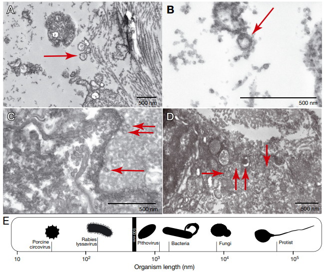
PSS was first identified in the 1950s, in a fish research center in Honolulu, Hawaii. Since then, there have only been 9 publications in the scientific literature documenting the condition and possible causes, although the fish community has come to the conclusion that PSS is likely a transmittable condition with an infectious agent as the cause. But despite this conclusion, there’s been no success so far in identifying such a cause – tests for parasites, bacterial growth, and viruses have come up empty-handed. That was until a 2021 paper, using high-resolution electron microscopy, found evidence of viral particles in facial tissues taken from Pacific mackerel. Suddenly, there was a lead: could PSS be caused by a virus that we just don’t have a test for yet?
Electron microscopy images showing viral-like particles (red arrows) in facial tissue from Pacific mackerel (Miller et al 2022).
Putting Together the Pieces
To investigate this hypothesis, this week’s guest Savanah Leidholt (a co-author of the 2021 microscopy study) is using an approach for viral detection known as metatranscriptomics. Leidholt, a fourth year PhD candidate in the Microbiology department, sees this complex approach as a sort of puzzle: “Your sample of RNA has, say, 10 giant jigsaw puzzles in it. But the individual puzzles might not be complete, and the pieces might fit into multiple places, so your job is to reassemble the pieces into the puzzles in a way that gives you a better picture of your story.”
Savanah Leidholt, PhD candidate in Rebecca Vega-Thurber’s lab, is looking for evidence of viruses in the tissues of fish with puffy snout syndrome.
RNA, or ribonucleic acid, is a nucleic acid similar to DNA found in all living organisms, But where DNA is like a blueprint – providing the code that makes you, you; RNA is more like the assembly manual. When a gene is expressed (meaning the corresponding protein is manufactured), the double-stranded DNA is unwound and the information is transcribed into a molecule called messenger RNA. This single-stranded mRNA is now a copy of the gene that can be translated into protein. The process of writing an mRNA copy of the DNA blueprint is called transcription, and these mRNA molecules are the target of this metatranscriptomics approach, with the prefix “meta” meaning all of the RNA in a sample (both the fish RNA and the potential viral RNA, in this case) and the suffix “omics” just referring to the fact that this approach happens on a large scale (ALL of the RNA, not just a single gene, is sequenced here!) When mRNA is sequenced in this manner, the researchers can then conclude that the gene it corresponds to was being expressed in the fish at the time the sample was collected.
The process of transcription: making messenger RNA from DNA. Image from Nature Education.
So far, Leidholt has identified some specific genes in fish that tend to be much more abundant in fish from captive settings versus those found in the wild. Could these genes be related to why PSS is only seen in fish in captivity? It’s likely – the genes identified are immune markers, and the upregulation of immune markers is well-known to be associated with chronic stress. Think about a college student during finals week – stress is high after a long semester, maybe they’ve been studying until late in the night and not eating or sleeping well, consuming more alcohol than is recommended. And then suddenly, on the day of the test, they’re stuck in bed with the flu or a cold. The same thing can happen to fish (well, maybe not the part where they take a test!,) especially in captivity – Pacific mackerel, tuna, and other scombrid species susceptible to PSS are fairly large, sometimes swimming hundreds of miles in a single day in the ocean. But in captivity, they are often in very small tanks, constantly swimming in constrained circles. They’re not exposed to the same diversity of other fish, plankton, prey, and landscape as they would be in the wild. “Captivity is a great place to be if you’re a pathogen, but not great if you’re a fish”, says Leidholt.
The results of Leidholt’s study are an exciting step forward in the field of PSS research, as one of the biggest challenges currently facing aquaculture farms and aquariums is that there is no way to screen for PSS in healthy fish before symptoms begin to show. Finding these marker genes that appear in fish that could later on develop PSS means that in the future a test could be developed. If vulnerable fish could be identified and removed from the population before they begin to show symptoms and spread the condition, then it would mean fish farmers no longer have to cull the entire tank when PSS is noticed.
The elusive virus
One of the challenges that remains is going beyond the identification of genes in the fish and beginning to identify viruses in the samples. Viruses, which are small entities made up of a DNA or RNA core and a protective protein coating, are thought to be the most abundant biological entities on the planet Earth – and the smallest in terms of size. They usually get a bit of a bad reputation due to their association with diseases in humans and other animals, but there are also viruses that play important positive roles in their ecosystems – bacteriophages, for example, are viruses that infect bacteria. In humans, bacteriophages can attack and invade pathogenic or antibiotic-resistance bacteria like E. coli or S. aureus (for more information on phages and how they are actually studied as a potential therapy for infections, check out this November 2021 interview with Miriam Lipton!) Across the entire planet there are estimated to be between 10^7 to 10^9 distinct viral species – that’s between 10 million and 10 billion different species. And fish are thought to host more viruses than any other vertebrate species. Because of technological advancements, these viral species have only really been identified very recently, and identification still poses a significant challenge.
As a group, viruses are very diverse, so one of the challenges is finding a reliable way to identify them in a given sample. For bacteria, researchers can use a marker gene called the 16S rRNA gene – this gene is found in every single bacterial cell, making it universal, but it also has a region of variability. This region of variability allows for identification of different strains of bacteria. “Nothing like 16S exists for viruses,” Leidholt says. “Intense sequencing methods have to be used to capture them in a given sample.” The metatranscriptomic methods that Leidholt is using should allow her to capture elusive viruses by taking a scorched earth approach – targeting and sequencing any little bit of RNA in the sample at all, and trying to match up that RNA to a virus.
To learn more about Savanah’s research on puffy snout syndrome, her journey to Oregon State, and the amazing outreach she’s doing with high school students in the Microbiology Department, tune in to Inspiration Dissemination on Sunday, November 20th at 7 PM Pacific!