
Choosing a subject for a protein portrait
This week we will put on our artist caps and earnestly search the protein data bank for subjects to portray. There are well over 100,000 structures on deposit in the PDB. How to choose just one?
Our interests in the science of proteins will strongly guide our choice of subject. Most of us have encountered topics in the life sciences that have piqued our curiosity and that we have told ourselves we will study further someday. Perhaps that day is here. You have seven weeks ahead of you to put together a protein portrait for public display. So if you are still curious about that nervous system twitch or that flower pollen antigen you heard about in Biology 211, that may be all the incentive you need to plunge into a deeper examination of the proteins associated with those fondly remembered topics.
 The search for a protein subject is also closely intertwined with our preferences for artistic media. Ask yourself in which artistic medium you are talented. And if the word “talent” is an overstatement, then ask yourself which medium you would like to play around in for the next several weeks. If you are a painter, say, you may look for a certain sort of protein subject, such as one that looks good in the gaudy colors of Calder’s Spiral Flowers.
Or if you have talent as a sculptor you may look for a different sort of subject, one whose molecular articulations are well presented by wiring and bended metals, like Calder’s Vertical Constellation. 
Or if you skipped art class in high school you may at least have stashed in the back of your closet a set of Leggo building blocks or an embroidery kit someone gave you for your birthday one year — Put that stuff to use by turning it into a protein portrait for 2016!
But an equally important source of inspiration is the simple tug we may feel when we encounter a protein while thumbing through the PDB. Check out this recently deposited structure, portrayed below by standard PDB thumbnail graphics. Without even knowing the name of the structure or its biological function, are you not intrigued by its pinwheel shape? Does the appearance of the molecule take you in an artistic direction? If so, go with the wind!

Click on the structure to go to its PDB page
Zeroing in on a protein structure in the PDB
Let’s look up one of the enzyme molecules that many people are interested in these days, HMG CoA reductase. This is the enzyme that is inhibited by the statin drugs, the drugs that block cholesterol synthesis. One of the most informative examples of the structure of HMG CoA reductase is listed under the code name 1hwk in the PDB. How did I discover this enzyme molecule in the PDB? I used the dynamic duo of online resources that every protein artist will use often:
- Good ol’ Wikipedia (I looked up the term “statin drug” and discovered an image of HMG CoA reductase which referred to the codename 1hwk), and then I used the amazing …
- Protein Data Bank (I searched for “1hwk”, or I could haves searched directly for “HMG CoA reductase”).

Once you arrive at the PDB entry for 1hwk, you can use sidebar commands to view the structure in 3D. You can change the appearance of the molecule. Change from view the proteins chains as ribbons to viewing space filling versions with every atom depicted by color schemes of your choice. There are a few varieties of 3D viewers to choose among. A lot depend on which platform and operating system you are using, be it a phone, laptop or console computer. Invest some time to explore.
Protein domains
Domains are compact arrangements of folded chains. From a purely artistic perspective, you can think of a domain as a major substructure (a chunk) of the overall protein. A domain stands apart from the rest of the structure. If a protein were a human body, the head would be one of its domains, the trunk another, the left arm another, etc. Some proteins have a single domain, others have many. A chain sometimes folds into a single domain , sometimes into multiple domains. Myoglobin is a single chain and a single domain. An IgG molecule includes four chains folded into six domains.
One of the great discoveries of the past decade is the conservation of domains across all of biology. The biological world includes a few hundred domains as the canonical elements that account for the structures and functions of essentially all of the many thousands of existing proteins. Long ago nature evidently discovered a set of compact machines (domains) and has used them creatively in assorted mix-and-match combinations. This is amazing: The many thousands of known protein structures (they all can be looked up in the PDB!) fold into just a few hundred generic protein domains.
For the artist, one very helpful exercise is to make 2-D sketches of 3-D proteins. A 2-D “topological diagram” can serve as a quick and easy surrogate for a rotatable 3D computer model when you are trying to make sense of how a chain travels through a molecule. Below are examples from Jane Richardson. She is deservedly credited as the inventor of 2-D topological sketches of proteins. Note how her topo diagrams readily highlight the differences between superficially similar alpha-beta class proteins:
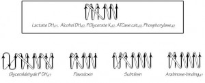
An artist can take advantage of 2-D topologies while attempting to depict the complicated multi-domain and oligomeric-structure of big protein molecules. No matter which depiction the artist has in mind, it is useful to make some 2D top0 sketches: Will you depict a small protein by showing its details, or will you smudge out the detail and portray a big protein? The sketch book is your friend!