
Begining with computer-rendered models of our favorite proteins available via the Protein Data Bank (PDB) and/or Alph Fold, we constructed new and highly individualized depictions. We chose shapes, colors, materials and media to tie the structures of the proteins to their function, bringing them to life.
We set up our show in the public area of the LINC building where honors college students were coming and going for finals, giving them a chance to aesthetically experience our creations during their study breaks.
Here is the 2025 parade of proteins portrayed by our fourteen student artists.
1. Rhodopsin
Artist: Ana Tracy
How do we see? In a word, light. You see that flower because rays of light scatter off it and deflect onto the lens of your eye, which focuses those rays onto your retina. Your retina records the focused image, much like a digital sensor in a camera. But how exactly does light hitting your retina translate into your conscious perception of the visual world?

PDB ID: 1F88 (Crystal structure of bovine rhodopsin)
Enter rhodopsin. Also known as “visual purple,” rhodopsin is a key photopigment expressed exclusively in rod photoreceptor cells of the retina. This protein consists of a small light-sensitive molecule of retinal (shown in pink in the right image below), bound inside the protein opsin (shown in orange and green). When retinal absorbs a photon, it changes shape, with one bond flipping from a bent (cis) conformation to an extended (trans) conformation (the right image below shows the trans, or activated, conformation). This tiny molecular signal is then captured by the surrounding opsin protein and used to launch a signal through a G-protein signaling network, which amplifies the signal and ultimately leads to a nerve signal to the brain.
 This protein portrait showcases the all-important covalent bond between retinal (light green) and lysine 296 of rhodopsin (dark green). Pulling on the yellow string simulates absorption of a photon by retinal, inducing the cis-trans flip of the bond. As retinal rotates into the trans position, it tips dominos representative of the signaling cascade which triggers our conscious perception of whatever is shining into our eyes – in this case, a floral arrangement revealed when dominos knock down the tented black cardstock in front of it.
The dominos are inscribed with a line from the poem “Mad Girl’s Love Song” by Sylvia Plath:
I shut my eyes and all the world drops dead;
I lift my lids and all is born again.

2.Icy InaZyme
Artist: Matthew Williams
While not technically an enzyme, InaA is a very cool ice nucleating protein found in the bacteria, Pseudomonas syringae.

The InaZ repeat sequence and the model InaZ9R construct Based on… ResearchGate. https://www.researchgate.net/figure/The-InaZ-repeat-sequence-and-the-model-InaZ9R-construct-Based-on-current-models-the-blue_fig1_349455179. Retrieved 4 June 2024.
Ice nucleation proteins (INPs) are capable of seeding ice crystals at freezing temperatures. They can be used to damage the cell walls of plant cells to gain valuable nutrients for growth or to seed precipitation to prevent residence in the atmosphere. Represented here is the slightly rectangular 𝜷-helix containing the ice-nucleation domain where ice crystals form at the planar repeats. The main helix is made from coat-hanger wire and the crystals are made from ammonium dihydrogen phosphate with a very small amount of blue coloring so as to appear like ice crystals emerging from the ice nucleation domain.

3. The Fading Mind. 40-Residue Beta-Amyloid Fibril
Artist: Jaymie Spano
PDB ID: 2M4J
 The 40-residue beta amyloid protein is found in the brain of those with Alzheimer’s Disease (AD). Beta-amyloid is a peptide that aggregates into long fibrils that accumulate and are observed in autopsies as vast depositions of plaques scattered amongst brain cells.
This material is thought by many to contribute to the development of Alzheimer’s Disease. As stated by the scientists who determined the molecular structure shown to the left state, “fibrils in the brain may spread from a single nucleation site, [and] structural variations may correlate with variations in AD.”
My protein portrait represents the unraveling of the mind.

I have integrated the basic parts of the 40-residue beta amyloid fibrils into a representation of how this molecular syndrome might afflict someone who could be one of my relatives or friends.
I placed each piece of the fibril in different areas to enhance the idea of plaques taking over the brain.
As the disease progresses, memories fade, like old photographs turning dark with time.
4. Green Fluorescent Protein (GFP)
Artist Name: Nathan Palazzolo
PDB ID: 1EMG and 1GFL
GFP is a protein heavily used for gene expression markers. Originally found in the Aequorea Victoriabioluminescent jellyfish to help glow under blue light, GFP has been extensively researched on its functions and applications. The fluorescence of GFP happens when a stimuli or excitation effect occurs, and a proton originally bonded to the Tyrosine-66 part of the chromophore in the middle of the cylindric GFP (pictured below) transfers over to the Glycine side.  The main body of GFP consists of 11 beta sheets that are bonded together with an alpha helix structure on the inside of the structure. There are also helical sections at the ends of the beta sheets that form the unique endpoints shown. Some research being done at Oregon State University involving GFP is genetic code expansion, where GFP is being used to confirm the incorporation of non-canonical (synthetically made) amino acids. When the non-canonical amino acids have been properly incorporated into the peptide chain, GFP will fluoresce and allow for the research to progress.
In order to represent GFP, I used LED lights and green foam sheets to represent how the beta sheets fluoresce when excited.  I also chose to focus mainly on the helical structure using the glass tube with the other elements wrapped around it. My original plan was to use copper tape and diodes to represent the fluorescence of the protein, but this quickly fell through when I realized that foam can be flammable when touching copper tape for long periods of time. Finally, in addition to providing a helpful stand for the structure, the soccer ball also resembles the head of a jellyfish when the structure is looked at upside down.
5. Voltage-Gated Sodium Channel
Artist: Anna Nielsen
Salt – tasty and exciting!

PDB ID: 7DTC
Salt has the chemical formula sodium chloride (NaCl). One of the ways the body uses sodium, Na+, is for transferring signals in the brain. A channel important to this pathway is the voltage-gated sodium channel. This channel is a protein! The channel opens in response to depolarization and allows sodium ions to flow into the neuron, as an action potential.

To represent the important role of voltage-gated sodium channels in excitability my protein art is bursting with colored pipe cleaners. The salt box standing for my protein is an ode to the sodium ions that flow through this miraculous channel.
6. Defender of the Brain: The Merlin Tumor Suppressor Protein
Artist: Kate Looney

PDB ID: IH4R
“Merlin,” short for “Moesin-Ezrin-Radixin-Like Protein” or “schwannomin,” is a cytoskeletal protein responsible for tumor suppression in humans. The protein consists of two large subunits, each containing several beta sheets and alpha helices, measuring about 70 kDa in total. Merlin is accountable for tumor suppression in neurofibromatosis type 2, a class of disorders characterized by their development of central nervous system tumors. In these disorders, the NF2 gene that encodes for Merlin encounters loss of function mutations or deletions, causing Merlin inactivation and resulting in nervous system tumors like meningiomas. Inactivation of this seemingly small protein can have catastrophic consequences. I experienced the ramifications of these mutations as I watched my own mother slowly, yet suddenly, develop into someone I had never met. Unbeknownst to us, a meningioma on her frontal lobe had grown to the size of a baseball, the extreme pressure causing loss of memory, personality, and motor control of her left side. Fortunately, excellent surgical practice ensured successful tumor removal, and my mother is now recovering at an incredibly fast rate. But as I watched her get wheeled off to the operating room, it was hard to fathom how a small protein dysfunction could catalyze so much damage.
In the brain model, I attempted to portray the pressure of the meningioma tumor by shaping the model asymmetrically with a divot in the front right lobe. The shape illustrates the force the tumor applied, and its power in changing the entire shape of the brain. The brain was made using cardboard for the base, hot glue for the folds, and paper mache for the texture.  A simplified model of the Merlin protein was made using pipe cleaners, oriented in the divot and projecting out of the brain in the way the tumor was. The contrasting colors indicate the two subunits, the purple and yellow colors representing my mother and my favorite colors, respectively. The protein is portrayed disproportionately large, illustrating both the magnitude of its importance and the consequences of its dysfunction.
7. Creating Bone with Cartilage Oligomeric Matrix Protein
Artist name: Caroline Lee
PDB ID: 1VDF
Cartilage oligomeric matrix protein (COMP) is a pentameric bundle comprising five right-handed alpha helices that coil around each other in a left-handed superhelix. 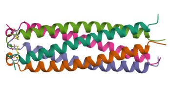 It is a non-collagenous extracellular matrix glycoprotein present in mammals, and found primarily within the human skeletal system, including articular cartilage, meniscus, ligaments, tendons, and synovium. COMP plays a crucial role in early bone and joint formation by facilitating collagen secretion and assembly, thus maintaining extracellular matrix stability. Mutations in the COMP gene can disrupt cartilage and bone formation, leading to pseudoachondroplasia and multiple epiphyseal dysplasia. These diseases result in incomplete bone growth, stunting an individual’s growth, and can cause early-onset osteoarthritis.
In crafting my protein portrait, I sought to preserve the protein’s structure while highlighting its role in bone formation and the consequences of when a mutation occurs.
 To achieve this, I envisioned the protein as the diaphysis of a long bone, reflecting its elongated and cylindrical shape, but kept it short to illustrate how a mutation can cause stunted growth. To highlight its coiled-coil structure, I employed distinct colors to improve visibility and underscore the presence of its five coils. Finally, to fully depict a bone’s anatomy, I included the epiphyses at both ends of the structure.
8. Voltage-Gated Potassium Ion (K+) Channel
Artist: Zach Fisher
PDB ID: 2A79
Potassium channels are a class of voltage-gated ion channel that open or close depending on the nearby electrical membrane potential. Voltage-gated potassium channels are the largest and most diverse class of such proteins, and serve a huge variety of functions.

In the axon of nerve cells, voltage-gated potassium channels are one of the three proteins responsible for generating an action potential: The neurological signal that travels through nerve cells, allowing them to communicate. Voltage-gated ion channels generate such a signal by allowing ions to flow into and out of the cell in response to elevated voltage (membrane potential) upstream. Thereby generating elevated voltage at their own position, and propagating the signal. Which travels down an axon like a wave.
My piece, a computer flight-simulation game, was inspired by our optional theme: artificial intelligence, and by a superb paper: Adaptive Flight Control With Living Neuronal Networks on Microelectrode Arrays, by Thomas B. DeMarse and Karl P. Dockendorf. Wherein DeMarse and Dockendorf grew neurons in a tissue culture dish lined with an array of 60 electrodes, and trained them to fly an F-22 Raptor plane in Xplane, a flight simulation program. I dedicate my game to the research animals, of the species Rattus norvegicus, involved in this research and similar studies.

Point your phone to the QR to join the action …

The only way forward for the potassium ion (the jet plane) is through the green channel proteins — use steady mouse control!
9. Uromodulin, The Immune System Mystery
Artist: Logan Kulisch
PDB ID: 4WRN
Uromodulin is a protein produced by the kidneys that is thought to have roles in regulating salt transport, protecting against kidney stones and urinary tract infections, and is thought to play a role in innate immunity in mammals.

I portrayed the protein using colored kidney beans adhered together with hot glue and mod podge, to represent the role uromodulin plays as a protein produced by the kidneys, and how it is essential for proper kidney function in mammals.

10. A Pig-ture Perfect Protein
Artist: Isabelle James
PDB ID: 1DZM
 The porcine odorant binding protein helps pigs have their un-boar-lievable sense of smell. It binds to odorants and transports them to receptors. The most interesting aspect of this protein is its distinct alpha helix and beta sheets configured in a spiral pattern. It almost looks like a snout, which was my inspiration for my protein portrait! Luckily, despite having an oink-credible sense of smell, hogs donít seem to mind their own odor.

11. Phage Attacks!
Artist: Lili Harris
PDB ID: 1AM7
The lambda bacteriophage is a small virus that infiltrates E. coli. It is reminiscent of the space shuttle landing on the moon, bringing foreign biology with it. Lysozyme is one of the first enzymes related to that invasion. It breaks apart the extracellular matrix of the E. coli, the sugar polymers that surround the cell for protection and interaction with the outside world. It is made up of three small homologous monomers:

In this sculpture, the lysozyme is represented with the helices and sheets painted on to resemble some sort of fur of an alien creature. A cleft is represented with white teeth, and the catalytic glutamic acid is highlighted with an eye. Here, it is breaking through the sugars represented with wire, which doubles as a structural support. The cell surface is represented simply, to look like a bumpy lunar surface.

12. Winnie’s Walks
Artist: Megan Easterday
PDB ID: 4ODD
 Can f 4 is a protein found in dog saliva and is one of the allergens associated with dog allergies. It is a lipocalin – a small, soluble protein involved in transporting small, hydrophobic molecules. Allergens like Can f 4 can trigger allergic reactions in sensitive individuals, leading to symptoms such as sneezing, itching, and respiratory distress.
My portrait depicts the amino acid sequence as Winnie’s leash, with each bead correlating to a specific amino acid, and Winnie, a clay golden retriever modeled after my own Winnie, a real golden retriever, who carries can f 4 with her every day!

13. Coagulation Cascade
Artist: Elana Crook
PDB ID: 2A1D
 Similar to legos or a lock and key, staphylocoagulase and prothrombin bind tightly together in the bloodstream to initiate the blood clotting cascade. Staphylocoagulase is a protein secreted by the pathogenic bacteria, Staphylococcus aureus. Infection of S.aureus is hallmarked by the increased activation of blood clotting via prothrombin – forming large clots on valves. The use of staphylocoagulase to activate blood clotting by binding to prothrombin helps S.aureus to bypass the host immune defenses, making it a more effective pathogen.
Within my artwork, staphylocoagulase is portrayed using orange beads while prothrombin is portrayed with red beads to demonstrate that these two proteins have a tight relationship when bound, but are individual from each other.  The tight structure that allows staphylocoagulase to bind to prothrombin is shown by the large number of alpha-helices, or spirals, within the molecular structure. The use of free beads on both staphylocoagulase (orange) and prothrombin (red) represent the amino acids that make up the individual molecular structures. The strings of beads on prothrombin (red) represent not only the long sheets of amino acids that make up its structure, but also the coagulation of blood as it clots during the binding of these two proteins.
14. Damage Suppressor Protein(DSUP)
Artist: Charles Axford
DSUP is found in tardigrades and it binds to and then protects their DNA from X-ray radiation. It is known as an intrinsically disordered protein which means it doesn’t have a consistent structure. Here is the Alpha Fold Predicted Structure:

Even AI is stumped! I conclude that little is known about what the majority of the protein looks like except for a pair of alpha helixes in the center, a common trait for proteins that bind to DNA.
So that gives me a lot of artistic freedom in my portrayal of this radiation-protective protein:

