Hello Everyone,
I will not be graduating until early 2018, so this is not my last post as an Oregon Sea Grant Scholar. However, this IS my last post as a Robert E. Malouf Scholar. With that in mind, I decided I would give a taste of what I have been doing this summer, but leave the “whole shebang” for my final post.
Recall, my project is characterizing metformin, a pharmaceutical drug, as a contaminant of emerging concern (CEC) in the lower Columbia River estuary. Fully characterizing a CEC in the environment should obviously include an ecosystem component. Thanks to the Malouf scholarship, I am currently exploring the effects of metformin on the lower food web.
This summer is best described as a series of never-ending lab experiments and instrumentation. It turns out that testing the effect of metformin on phytoplankton and microbes is harder than it looks! Currently, my lab bench looks like an explosion of flasks, vials, pipettes, and sediment. Like most scientists, I spend more of my time washing and prepping experiments than I do actually collecting data – an unromantic fact of the scientific existence that the public tends to ignore. I will focus on my phytoplankton work, since the microbe experiments are currently taking an unexpected turn (more on that in the future!).
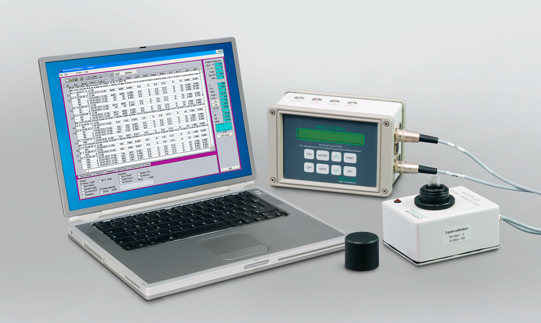
Most of my phytoplankton work has revolved around the PAM fluorometer – an awesome device that measures the kinetics of fluorescence in photosynthetic cells.

The simple beauty of PAM.
If you recall basic photobiology, photosynthesis (the production of energy from sunlight) works through an electron transport chain that takes place in the thylakoid membrane of chloroplasts in photosynthetic cells. Pigment molecules (e.g. chlorophyll b, xanthophylls, or carotenes) within a membrane photosystem (Photosystem II) absorbs photon energy from sunlight. This energy is used to excite electrons donated by oxygen in water. The excited electrons are held less strongly by the oxygen nucleus and escape into a “pigment funnel” (i.e. antenna complex), where it is passed from pigment molecule to pigment molecule toward an ultimate electron acceptor (i.e. chlorophyll a) at the reaction center of the photosystem. The chain continues after the electron is shuttled to another photosystem (Photosystem I) and a different electron acceptor. The electron will ultimately be used in the production of NADPH and a proton gradient that fuels ATP synthase to produce energy.

The concept of photosynthesis: an electron from water is excited to a higher energy state and escapes into Photosystem II where it is passed from pigment molecule to pigment molecule until it hits an ultimate electron acceptor. The electron is then passed through an intermembrane system to Photosystem I where it is funneled to another electron acceptor and eventually used to reduce NADP+ to NADPH. This forms an intermembrane proton gradient that drives ATP synthase and the production of energy.
In a nutshell, the PAM fluorometer stimulates and measures this process by delivering light pulses to a water sample containing photosynthetic cells (algae, in my case). Recall, photosynthesis is based on the idea of an excited electron losing energy. Whenever an excited electron moves to a lower energy state, it must release energy. This energy is released in the form of long wavelength fluorescence, which can be measured by a fluorometer. By measuring minimum and maximum fluorescence in response to a series of light pulses, the PAM fluorometer can measure electron transport rate and non-photosynthetic quenching processes (e.g. energy lost via heat, instead of fluorescence). In my case, this is perfect for looking at the effect of a chemical compound on photosynthetic efficiency.
My labmate jokingly dubbed the PAM fluorometer a “magic box”, however, there is a scary amount of truth to that term. The PAM fluorometer is an amazingly compact device that can measure an amazing number of photosynthetic processes. People tend to take it for granted since it appears simplistic: turn it on, insert sample, measure photosynthesis, and you’re done! WRONG.
There are so many quirks to the device. First off, the manual. Or, should I say, manuals. Part of one topic will be explained in one manual, but an important note on the same topic will be explained in the other manual. Confusing is an understatement. Let’s not mention the calibration protocol which cost me a month of fiddling.
As a result, I have learned the PAM fluorometer from the inside out. Some examples of what I have learned over the past three months: (1) there is an optimum time to “dark-adapt” samples to make sure photosystems are completely receptive to light (i.e. completely empty of electrons), (2) far-red light should also be used to fully oxidize Photosystem I and the intersystem electron transport chain to maximize electron receptivity, (3) the sample should be diluted until the fluorescence value does not exceed a maximum value, and (4) the sample cuvette has an optimum volume and should be cleaned with ethanol in between samples to prevent obscured light paths. These are just some of the many things that I had to learn by trial and error through countless failed phytoplankton experiments.
I am aware that this entire post is about the PAM fluorometer, but it has arguably been my greatest achievement this summer. I am proud that I am mastering such a deceptively complicated device. By the end of this project, I plan on having written a detailed calibration protocol and detailed illustration of the PAM fluorometer so that future lab members can easily take measurements without a similar summer of trial and error. The long process was a blessing in disguise though – I truly have a better grasp on photosynthesis and fluorometry instrumentation.
What about the results? Well, if you don’t make it to the CERF conference this year, you will have to wait for my last blog post. I am currently doing my fine-tuned toxicity experiments with Chlorella vulgaris (a basic green algae) and will be performing the same experiments with Thalassiosira weissflogii (a diatom, which tends to be more sensitive). This should give us a good idea of the effect of metformin on the photosynthetic processes of two representative organisms in the Columbia River estuary.
Stay tuned.


