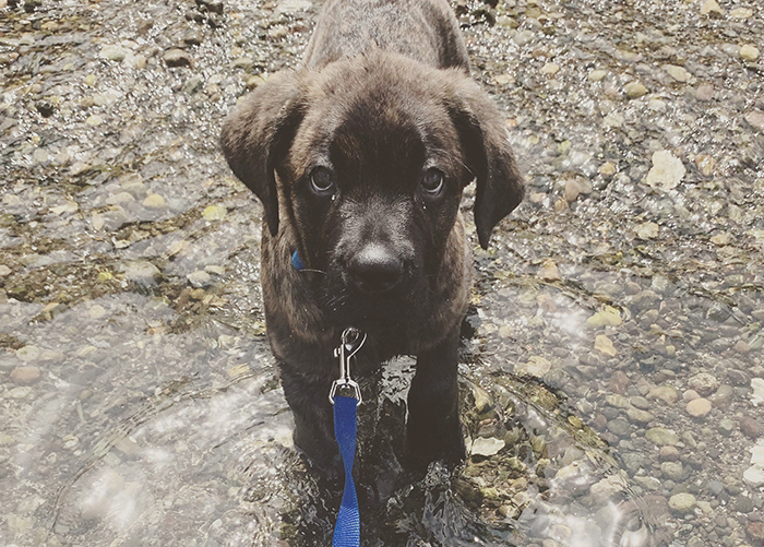
Breeds like bulldogs and Pekingese are called brachycephalic dogs; they have skull bones that are shortened in length, giving the face a pushed-in appearance. This characteristic of their anatomy can cause health problems, including respiratory conditions.

Brachycephalic airway syndrome (BAS) refers to a set of upper airway abnormalities that sometimes affect brachycephalic dogs. Mildly affected dogs will have noisy breathing, especially with exercise. Severely affected dogs have more pronounced airway noise, get tired more easily, and may faint after exercise.

In December, a pug was referred to the OSU Veterinary Teaching Hospital (VTH) with shortness of breath. Radiographs showed multiple airway abnormalities, typical of brachycephalic airway syndrome. The owners met with Dr. Lea Mehrkens, a soft tissue surgery resident, to discuss treatment options. They decided to proceed with corrective surgery.

Dr. Katy Townsend, a soft tissue surgeon at the VTH, increased the diameter of the pug’s nostrils, shortened the soft palate, and removed obstructions in the larynx. The pug was then admitted to the Intensive Care Unit (ICU).

Dr. Tandi Ngwenyama is head of the critical care team in the ICU. She works with a group of interns, residents, and highly-skilled veterinary technicians to monitor and care for patients recovering from surgery, or with other life-threatening conditions. 

The ICU team closely monitored the patient during her recovery from surgery and noticed she was still having trouble breathing due to post-surgery swelling. Dr. Ngweyama consulted with the surgeon, discussed different options, and decided to proceed with a temporary tracheostomy.

Back in ICU, following the tracheostomy, Dr. Ngwenyama developed a new plan for the pug’s care. It included continuous checking for airway and tube obstructions, and monitoring for comfort.

 “She spent three weeks with us and we got very attached to her. One of our goals is to make the ICU as positive an experience as we can for the patient. She is a sweet, loving dog so we were very happy that the outcome was good.”

Dr. Ngwenyama is new to the critical care service at the VTH. “I am excited to join the leadership team,” she says. “Technician supervisor Julie Posch has done an amazing job; we have similar philosophies and vision for the ICU.”

One of Dr. Ngwenyama’s first objectives was to work with the ICU team to write a new mission statement:  The SA ICU team demonstrates compassionate care, and strives for excellent patient care while fostering a positive teaching and learning environment for all faculty, staff and students.

“Now we want to make sure that we live up to it; that our actions, thoughts and words are consistent with what are values are,” says Dr. Ngwenyama. “We want to actually walk our talk.”

To that end, the group decided to make real changes that will improve communication, particularly in daily rounds. “Because we work 24/7 and have shift changes,” says Dr. Ngwenyama, “rounds are very important for communicating with the next group of people. That is when we discuss the status of the patient and our goals for their care.” The ICU team is working on a more structured approach to rounds to make them more effective. “We want them to be clear, concise, and complete.”

Another goal for the ICU team is to add the practice of debriefing. “I think it is very important with high intensity cases, or if you, unfortunately, have a poor outcome,” says Dr. Ngwenyama. “We need to meet as a group, for even five or ten minutes, to debrief. For example, we had a CPR case and unfortunately could not get spontaneous return of circulation. There were a number of people and services involved: certified technicians, house officers, students, the anesthesia service and internal medicine. At the debrief, it was so great to meet and follow a structure: we did a quick synopsis, then talked about what did well, and what we could have done better.”

Dr. Ngwenyama also thinks it is important to create an environment where everyone can have a voice. “It needs to be a flattened hierarchy where not only doctors can speak,” she says. “Everyone’s perspective is important.”

The microbiome is the sum of all the bacteria that live in and on an organism. These bacteria can play an important role in keeping unwanted pathogens, such as viruses, from getting a foothold and causing disease.

Dr. Brianna Beechler is a researcher at the Carlson College of Veterinary Medicine. She has joined forces with Dr. Rhea Hanselmann, of the Western University of Health Sciences, to study how the microbiome in the respiratory tract differs between cats with and without upper respiratory infections.

Both researchers have a veterinary clinical background and know how devastating upper respiratory infections can be in cats. They have wondered why some cats are resistant to infection, even if they are sharing a home with an infected cat. “We don’t know why some animals can have a pathogen and exhibit clinical signs and others have the same pathogen and don’t,” said Dr. Beechler. “We say it’s related to stress, but what does that really mean? We know there’s a link in African buffalo between their upper respiratory microbiome and whether they get other diseases or not. We thought the same thing might be happening in cats.”

Faced with a rare lung disease plus a liver disease that was virtually unknown, it took a team effort to save Chance.

At twenty-seven Chance is an older horse, but he still enjoys going for a ride with his owner. Last winter Chance started losing weight and energy. A visit to his veterinarian resulted in several abnormal blood tests that made her concerned about his liver, so she referred him to the OSU Veterinary Teaching Hospital (VTH) for further evaluation.

Dr. Ana Pacheco is a large animal internal medicine specialist at VTH. She examined Chance, performed an ultrasound, and ordered a complete blood workup. The ultrasound showed an unexpected result: not only did his liver appear abnormal, but also his lungs. Dr. Pacheco ordered x-rays of his chest which revealed large nodules on his lungs. She had those biopsied and sent the tissue to the Oregon Veterinary Diagnostic Laboratory. The pathologist at the lab found severe inflammation and fibrosis of Chance’s lungs, and diagnosed Equine Multinodular Pulmonary Fibrosis (EMPF), in conjunction with equine herpesvirus. The big surprise: they also found herpes and fibrosis in his liver.

EMPF was first discovered in horses in 2007. Equine herpes in the liver is very rare. So Dr. Pacheco was faced with a lung disease that had only recently been studied, and a liver disease that was virtually unknown.

Dr. Karen Labbe, a veterinarian who was completing a fellowship at the VTH, was part of the team caring for Chance. She combed through recently published studies, and was able to find a couple of cases in an Australian journal where horses with EMPF also had liver fibrosis. With that information, she and Dr. Pacheco decided to start Chance on four weeks of steroids to bring down inflammation and slow the progress of the fibrosis. They also put him on a new diet of hay-free alfalfa feed.

Chance was a very sick horse when he arrived at the OSU Veterinary Teaching Hospital

The prognosis for EMPF treatment is fair to poor, but at his one-month checkup, Chance showed significant improvement in weight gain and body condition, and his liver tests were close to normal. His chest x-rays showed significant reduction of the fibrosis.

At his next check up, six months later, Chance showed even more improvement in both his lungs and liver. “Most important was his incredible improvement in attitude and body condition,’ says. Dr. Pacheco. “He was 145 pounds heavier than when he first came into the hospital. He made an amazing recovery.”

Dr. Pacheco recognizes that the team approach at the VTH is a big advantage in treating her patients. “We have so many opportunities to collaborate,” she says. “I can work with radiologists, and pathologists, and other specialists. We also have great equipment and great technicians. It’s a big group who are involved in caring for our patients.”

Chance is currently feeling good and enjoying a normal life. “He has slowly gotten better and better,” says his trainer, Tobey Spitzer. “I started working him slowly on the ground and now I am riding him. He looks beautiful!”

Every day the OSU Veterinary Teaching Hospital witnesses inspiring stories of the human-animal bond in clients who go to great lengths to ensure the good health of their pets. The Kendall family is one such story. In November, Garret, Patricia and their three kids gave their all, financially and emotionally, for a puppy named Griffey that they had only known a few months.

Griffey joined the Kendall household last summer when he was 6 weeks old. He is a Cane Corso big-breed dog, but was the runt of the litter. His joyful, gentle personality quickly captured the hearts of the Kendall family and everyone he met.

When the Kendalls took Griffey to his first checkup, their veterinarian heard a heart murmur, a common condition, generally not too worrisome. Several months later the murmur was worse, so they took Griffey to a cardiologist who found congenital deformities in the puppy’s heart, and referred them to OSU.

Dr. Nicole LeBlanc is a cardiologist at the Lois Bates Acheson Veterinary Teaching Hospital. She and resident Eric Owens evaluated and diagnosed Griffey with four different heart defects including a misplaced aorta, a hole between the lower chambers of the heart, and pulmonic stenosis (the valve between the heart and the lungs is too narrow).

Drs. LeBlanc and Owens knew surgery could help, if not cure, Griffey so they consulted with Drs. Katy Townsend and Milan Milovancev in the soft tissue surgery service at OSU. The two teams decided that the best way to help Griffey was to perform a rare procedure called a Blalock-Taussig-Thomas shunt, where a tube is grafted between the heart and lungs. “The shunt allows the de-oxygenated blood from the aorta to be transported to the pulmonary artery where it is circulated through the lungs and becomes oxygenated,” says Dr. Milovancev. “It is a palliative measure to decrease the severity of his condition.”

By the time the Kendalls met with Dr. Milovancev, Griffey’s condition had worsened and it was having a significant effect on his life: he got tired easily, was slow to get up and go outside, and was even laying down to eat.

Dr. Milovancev explained the challenges for the shunt surgery: he told them that it would not give Griffey a completely normal heart, but it would increase the oxygen in his blood significantly. He told them the prognosis was good for Griffey’s return to normal physical activity, once he had recovered from surgery.

Despite the risk and considerable cost, the Kendalls elected to move forward. “The decision wasn’t easy,” says Garrett Kendall, “but we felt like we owed him a chance at a better heart because he gives us his whole heart every day. He is a full-fledged member of our family and our kids love him dearly.”

Another factor involved in the decision to proceed with the surgery: it had only been performed on a few dogs, and never at OSU, so it was an opportunity for the doctors to refine a life-saving skill. “I told Dr. Milovancev that good or bad, something positive would come from Griffey’s case. Our hope is that in the future they will be able to help other dogs and families.”

Drs. Milovancev and Townsend began a process of extensive planning for the graft surgery. “It was technically challenging and required special equipment,” says Milovancev. Planning up-front also saved time under anesthesia, which was safer for Griffey, and it minimized cost.

Milovancev was also able to save money by getting the manufacturer to donate the ‘tube’ being grafted in Griffey’s heart. “Normally it would cost the client about $1,000,” he says.

The surgery went very well. As Griffey recovered in the Intensive Care Unit, fourth-year student Alexandra Hoff facilitated communication between doctors, ICU, and Griffey’s owners. “I made twice-daily phone calls to update the owners on Griffey’s condition,” she says. “I performed daily physical exams, wrote his treatment sheets (checked by doctors), and most importantly, I made sure he had enough hugs and kisses.”

Being assigned to Griffey’s case was a unique learning experience that Hoff really values. She was responsible for his pre-op and post-op care, and was allowed to scrub-in and observe the surgery. “I was able to feel the blood moving through the shunt after it was placed, and the whole surgery was the coolest procedure I’ve ever been involved with.”

Her close, personal attention to Griffey also gave her an exciting look at his recovery. “It was very cool to see how quickly his cyanosis improved,” she says. “Prior to surgery, his gums were completely purple due to lack of sufficient oxygen; immediately post-op his gums were light pink in color.”

Griffey’s case gave Hoff the opportunity to learn about a segment of cardiology that few students study. “I reviewed the physiology of both left and right shunting cardiac anomalies, and the difference of the impact on the body between the two,” she says. “I read an excellent paper discussing the modified Blalock Taussig shunt; and I learned the important aspects of post-operative care in cardiac cases, and how to manage these cases in both pre- and post-operative periods. Overall it was an incredible experience and both Dr. Milovancev and Dr. Townsend were great!”

The Kendalls also appreciate the doctors and all the people involved in Griffey’s care. “We are so grateful for the work that they did,” says Garrett Kendall. “Our experience at OSU was incredible. Everyone we came into contact with showed a genuine care for Griffey and a love for what they do.”

Although still recovering from his surgery, Griffey’s oxygen levels will continue to improve over time. “He is full of energy,” says Kendall. “He loves to pounce around and play. We are excited to take him to the dog park when he is fully healed.”

From community outreach events to the classrooms of Magruder Hall, dogs can be found helping out.

The student-teaching room is lined with stainless steel tables, but step into the neurology class on exam day, and you will see students down on the floor with dogs. The dogs aren’t patients, they are ‘teaching assistants’ who work for hugs and treats.

Where does the college get these furry, cooperative teachers? Very often they are the pets of students and faculty, and the ones who crave lots of attention often participate in many different learning activities throughout the college.

Sophie and Pasco are instantly recognizable walking down the hallways of Magruder Hall. Sophie is a tall, regal standard poodle with impeccable grooming. Pasco is a tiny furball. They are well-known in the CCVM, not only in the classroom, but also for their participation in student events, and in a wide-variety of community outreach efforts. They belong to student Eilea Delgadillo.

“They are both old and well-socialized,” says Delgadillo. “I was a groomer before I went to vet school, so they are both very used to frequent handling.”


Sophie and Pasco have had their teeth cleaned by the dentistry class, and provided ultrasound images for veterinarians taking continuing education training. Dozens of elementary school children have used a stethoscope to hear their kindly canine hearts beating at Science Nights, and in programs like How We Role, which introduces kids to veterinary medicine with a goal of diversifying the profession.

Although students are only allowed to bring assistance dogs to school with them, the occasional ‘teaching’ dog is an exception. Student Nikita Neuhaus has an Australian Shepard mix named K-Dog who volunteers at the college regularly. “She has come in for almost every student teaching lesson we have had,” says Neuhaus. “She was the demo dog for physical exams, for body condition scoring, and for neurological exams. She has also very patiently allowed us to draw her blood.” Now that is really going above and beyond, but K-dog gets a lot in return.

“She loves attention. All she wants is for someone to pet her and snuggle her, so having a whole class full of people who do that is like a dream come true, even if it means a little poke.”

Students Gain More Than Knowledge From India Experience

Throughout their four years at OSU, veterinary students have the opportunity to participate in many externships, from a few days at the Portland Zoo to a few weeks at a local veterinary clinic. One of the most extraordinary externships provided by the CCVM is a month-long stay at the Karnataka Veterinary University (KVU) in Bangalore, India.

This year Donald Gridiron was among a small group of students selected to participate. His externship included hands-on training in internal medicine, obstetrics, and surgery, plus unique learning experiences like working with zoo veterinarians. “The most interesting case I worked on, by far, was a surgery at the Mysore Zoo. We got to observe and assist in removal of a mass from a seven-year-old tiger named Chamundi,” says Gridiron. “We acted as the anesthesiologists, monitored vitals, and collected blood.”

The Veterinary Teaching Hospital in Bangalore is government funded, and services are available at a low cost which makes it a very busy place. This gave the CCVM students a lot of experience in a short amount of time. “I spent just under three years in general practice before entering veterinary school, and saw two or three pyometra surgeries the whole time,” says Gridiron. “On the OB rotation [in India], I observed two or three a day.”

Another difference in the Bangalore teaching hospital: Small animal medicine is provided in one big room with many exam tables, holding a variety of animals, receiving a variety of treatments. “I got to see good examples of practicing medicine with what you have available and not necessarily what is considered the gold standard, and realizing that it still benefits the animal,” says student Sabrina Dean. “This knowledge will be important in future practice, because I will work with clients who cannot afford to do everything I want for a pet.”

Both Gridiron and Dean were actively involved in cases not available to second-year students in the U.S. “I got to assist with a traumatic abdominal hernia repair in a sheep,” says Gridiron. “The guts were literally hanging out . . . it took eight people to properly position the sheep and surgically close the tear in the muscles.”

That kind of hands-on participation is a confidence-builder for students just starting a difficult profession. “I gained confidence in myself by being there,” says Dean. “I really had to break out of my shell, and become responsible for making sure I was engaged and asking for what I needed, which will help me a lot when I start doing clinical work.”

The India Externship Training Program was conceived by Dr. Manoj Pastey, Associate Professor of Virology, to foster collaboration between CCVM and his alma mater. It is financed by the Department of Biomedical Sciences. All the students on the trip gained invaluable experience.

“This trip helped remind me why I love doing what I do,” says Gridiron. “Even though things may be different around the world, people still love their animals just the same, and to me that was the coolest part. By the end of the trip I was really able to understand the importance of a veterinarian in any society.”

Last year an Oregon woman found a baby squirrel and tried to nurse it back to health. Although she had the best of intentions, that turned out to be a bad decision. Not only did the squirrel die, it exposed the woman to Francisella tularensis, a bacteria can be lethal without treatment.

Fortunately, the woman sent the dead squirrel to the Oregon Veterinary Diagnostic Laboratory (OVDL) for evaluation. At the OVDL, a pathologist found suspicious lesions in tissue samples taken from the squirrel and forwarded them to the bacteriology department who identified Francisella tularensis. The OVDL  contacted, the owner and notified and sent samples to the Oregon State Public Health Laboratory (OSPHL) who confirmed the diagnosis. The OSPHL informed the exposed woman, and advised her to begin antibiotic treatment.

Although the OVDL often works closely with the OSPHL, and the outcome of this case was successful, it also served as a catalyst to bring the two agencies together for an analysis of the processes they use to respond to biological risks. As a result, they further refined a triage protocol for high-consequence pathogens that would provide added precautionary steps in the future.

As part of the new protocol, the OVDL modified their sample intake forms to better gather high-risk information. They added questions like What symptoms is the animal showing? and Was there exposure to humans?. The new forms will enhance safety in the laboratory and help prioritize cases.

The whole process was a successful collaboration between the OVDL, the OSPHL, and the Oregon Health Authority.

Thanks to the Olive K. Britt endowment, Brie received the surgery needed to go to her forever home.

When critically ill shelter animals come to the OSU Veterinary Teaching Hospital (VTH), part of the cost of their housing, surgery, and care is paid by the Olive K. Britt Endowment for Emergency Medicine.

Olive Britt was an OSU alumna who earned a degree in Wildlife Sciences in 1940, and went on to become a veterinarian. When she died in 2006, her will created an endowment to provide hospital care for shelter animals, and animals whose owners could not afford critical treatment.

Dr. Kirk Miller, an OSU faculty member teaching veterinary students at the Oregon Humane Society in Portland, has sent several cats and dogs to OSU for procedures that he cannot provide. “These are young, otherwise healthy animals that are very adoptable,” he says. In fact, he says, “They rarely make it back to us. They get adopted there [Corvallis].”

Last month, that happened once again when a homeless Golden Retriever named Brie was brought to the VTH for repair of an Ectopic Ureter. In normal dogs, the ureter connects the kidneys to the bladder. Brie was born with a ureter that did not connect to her bladder. The VTH surgically repaired this defect, and the Britt fund paid part of the cost. Soon after Brie recovered from surgery, one of the students working on her case found her a forever home with a friend in Portland, Callan Christman.

“We are so grateful for all that OSU has done to make Brie healthy,” says Christman. “She is a wonderful addition to our family and we love her so much.”

The Olive K. Britt endowment earns about $7,000 a year in interest. In 2019, donors contributed another $4,360. All that money has been used to help shelter animals and pets of low-income owners. The many, many generous donors who have contributed to the Britt fund over the years have enabled the VTH to save the lives of hundreds of beloved pets and pets-to-be.

If you would like to make a gift, large or small, to the Olive K. Britt endowment, visit the OSU Foundation website.

Before his accident Tucker could “jump four feet straight up, spin, and fly through the air to catch a ball,” says his mom Rhonda Reed. “He could also climb trees. He could run right up the trunk, and sit in the crotch of the tree ten feet up.”

Two years ago, on a rare snowy day in Eugene, Reed let Tucker out to enjoy the experience. He slipped on the ice and fell, but typical for him, he just bounced back up and joined his canine siblings in play.

The next day Tucker was limping, so Reed took him to her veterinarian who gave him pain medication and a steroid injection for inflammation. Unfortunately, over the weekend, Tucker got worse, but a trip to the emergency clinic for x-rays was inconclusive. By Monday, Tucker was completely paralyzed in both hind legs and in a lot of pain.

Reed frantically called several veterinary hospitals looking for help. Finally, she was referred to the Lois Bates Acheson Veterinary Teaching Hospital (VTH) at Oregon State University. With two board-certified orthopedic surgeons and a large orthopedic team, the VTH treats hundreds of dogs with disc injuries every year, so the staff answering phones know how important it is to act quickly in cases of paralysis. In Tucker’s case, they described his symptoms to Dr. Isaac Cortez, an orthopedic surgery resident, and he arranged to bring Tucker into the hospital immediately. There, an MRI confirmed Dr. Cortez’s suspicion of herniated discs that were extruding into the spinal canal and compressing Tucker’s spine.

The success of any disc surgery is dependent, in part, on relieving spinal compression quickly. Dr. Cortez and orthopedic surgeon Dr. Jennifer Warnock took Tucker into surgery that afternoon. They performed an extensive procedure, removing the protective roof of the spinal canal to access and remove bulging disc material between four vertebrae.

Soon after waking up, Tucker showed one small sign of improvement: he was able to wag his tail, but he still had a long road ahead to regain movement in his legs. Sara Short, a Certified Rehabilitation Technician began laser therapy on Tucker while he was still in ICU. “It is unusual for us to have a case with this many disc injuries,” she said, “so we wanted to promote healing and decrease inflammation right away.”

Continue reading

From hummingbirds to bears, there is often something interesting (and a bit gory) on the steel tables in the necropsy laboratory.


Stacie Nellor handed a stainless steel bowl up to the first person in the front row of the viewing gallery. In the bowl was a feline abdomen that contained a large tumor. The specimen was passed among a group of two dozen students while Nellor explained the source of the cat’s abdominal bleeding: a rare disease called Factor XI Deficiency.

Nellor is a fourth-year veterinary student working in the necropsy lab where, among other things, she performs animal autopsies (called necropsies) under the supervision of a pathologist. Nellor saved the cat’s remains to present at the Wednesday morning necropsy rounds. The rounds are open to any student or staff of the Carlson College of Veterinary Medicine, and sometimes visitors on guided tours attend as well.

The necropsy lab is one of the busiest services at the Oregon Veterinary Diagnostic Laboratory (OVDL). In addition to educating students, pathologists perform necropsies for veterinarians, pet owners, and farmers, and work closely with many government agencies from Oregon Public Health to the U.S. Department of Agriculture.

Dr. Rob Bildfell is an OSU professor, a board-certified pathologist, and supervisor of the necropsy service. In his twenty years at the lab, he has seen a wide range of interesting cases involving many different species, and a lot of those came from the Oregon Department of Fish and Wildlife (ODFW).

From big bears to tiny hummingbirds, ODFW is interested in knowing why animals have died. “Typically they send us species that are of interest to their clientele, like deer, elk, and ducks,” says Bildfell.  “Or they may be concerned about disease transmission when an animal has had contact with humans, like a wild rabbit that a Good Samaritan has tried to help.”

Continue reading